

Abstract
Objective: This study aimed to evaluate the survivability and success rate of medical trauma implants used in orthopedic trauma surgery. Fracture union rates, postoperative complications, and implant failures were analyzed to determine the overall performance of the implants.
Materials and Methods: The study included 455 patients (621 fractures) who underwent surgical fixation with Oltho Medical orthopedic trauma implants at Hacettepe University between January 2020 and July 2022. Patients with at least six months of follow-up and adequate radiographic evaluation were included. Fracture healing was assessed using the Radiographic Union Scale for Tibia (RUST) and a similar four-cortex scoring system for other fractures. Implant failures, postoperative complications, and patient demographics were recorded.
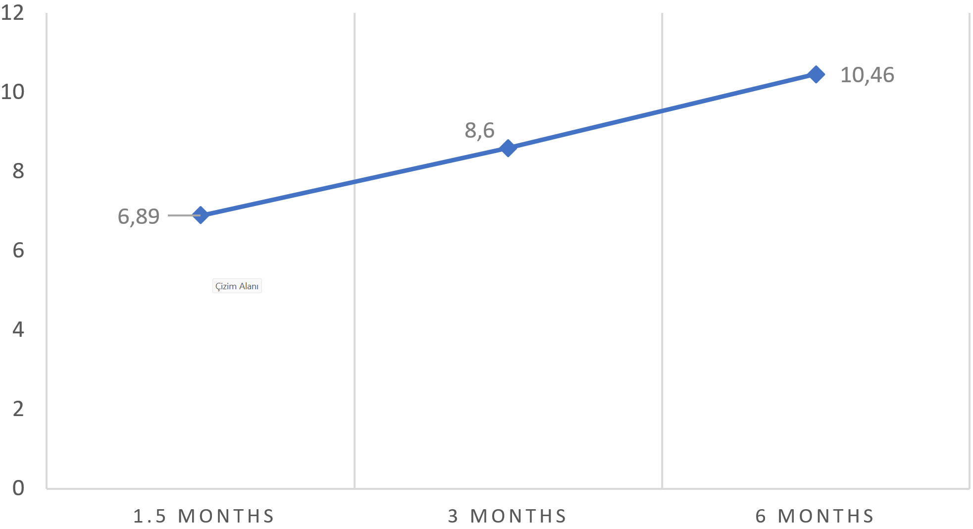
Results: The mean patient age was 34.2 ± 24.3 years (range: 6–93), with a mean follow-up of 16 months. The overall fracture union rate was 97.2% at six months, with a mean RUST score of 10.5. The implant survival rate was 98.7%, with implant failure observed in six (1.3%) cases. Postoperative complications occurred in 7.3% of patients, including wound complications (6.2%), nonunion (1.3%), and implant failures (1.3%). Although implant-specific survival rates varied among anatomical regions, all fractures ultimately achieved union following appropriate management.
Conclusion: Medical trauma implants demonstrated high survivability and effectiveness in fracture fixation, with a low failure rate comparable to existing literature. Despite some complications, all were successfully managed, supporting the reliability of these implants in orthopedic trauma surgery.
Keywords: orthopedic implants, fracture healing, trauma surgery, implant failure, survivability
Introduction
Bone is a dynamic tissue, and the damage that occurs after trauma initiates the healing process due to the high remodeling and regeneration capacity of the bone [1]. Fracture healing is a physiological process that is affected by biological and biomechanical factors [1,2]. Fracture stabilization and fixation are essential for bone healing, making orthopedic implants indispensable in trauma surgery [3,4]. Successful fracture healing depends on biological, mechanical, and biomechanical factors, as well as appropriate surgical management [5,6]. The path to be followed in the detection and treatment of the fracture is affected by factors such as trauma severity, fracture location, soft tissue condition, patient’s medical condition, bone quality, and surgical experience [7].
Fracture healing is a dynamic process influenced by multiple factors [3,8]. Bone healing occurs through two primary mechanisms: primary (direct) healing and secondary (indirect) healing. Primary (direct) healing is rare and occurs when absolute stability is achieved, with interfragmentary movement of less than 0.01 mm and strain below 2% [9]. Secondary (indirect) healing is the most seen type, and it is a form of recovery after fracture end movement and weight bearing. It is seen in all non-surgical treatments, also in intramedullary nailing, external fixation, and some internal fixation treatments in complicated comminuted fractures [10,11].
The design of orthopedic implants has shown certain changes from the time it was started to use [12]. Modern implants improve bone healing through improved biocompatibility, mechanical stability, and surface chemistry [12]. Implant-bone healing mechanism differs depending on implant surface chemistry and topography [13]. Peri-implant incompatibility causes complications as nonunion and multiple failure due to implant insufficiency [14,15]. Nonunion occurs in approximately 2% of all fractures but may reach 20% in diaphyseal fractures [15]. The U.S. Food and Drug Administration (FDA) defines nonunion as the absence of healing within nine months or lack of progression over three consecutive months [16].
Fracture healing is assessed through both radiological and clinical evaluation [17]. While pain during weight bearing is considered clinically significant, radiologically progressive improvement in serial radiographs and cortical bridging are also to be considered [18]. Healing scores in four cortices on anterior-posterior and lateral radiographs has been modified recently, taking into account the bony bridging and non-bridging callus tissue, that has been applied in certain fracture types [19,20].
This study aimed to assess the survivability and success rate of medical trauma implants in various surgical fixation methods across different fracture types. Additionally, we aimed to analyze patient demographics, comorbidities, fracture union rates, and associated complications to further evaluate implant performance.
Material and methods
This study was approved by the Institutional Ethics Committee (IRB No: GO 22/942). We retrospectively analyzed all patients who underwent surgical fixation with Oltho Medical orthopedic trauma implants (Ankara, Turkey) at Hacettepe University between January 2020 and July 2022. Routine postoperative follow-up was conducted at 2 weeks, 6 weeks, 3 months, 6 months, 1 year, and annually thereafter. The study included patients with at least 6 months of follow-up and proper antero-posterior and lateral views of the fracture site. Patients with less than 6 months of follow-up and inadequate radiographs were excluded from the study. A total of 601 patients were identified. Of these, 92 were excluded due to inadequate radiographs, and 54 were excluded for having less than 6 months of follow-up. Immediate postoperative, 3-month, and 6-month radiographs were analyzed for fracture union and implant failure (loosening, breakage, and subsidence). Fracture union was determined as viewing bridging callus formation on radiographs (3 out of 4 cortices in anteroposterior and lateral views if possible] and non-tender/painless palpation of the fracture site). Patient demographic data and clinical notes were retrospectively collected from medical records. Radiological evaluation was conducted independently by a fellowship-trained orthopedic trauma surgeon using the Picture Archiving and Communication System (PACS).
Tibial fracture healing was assessed using the Radiographic Union Scale for Tibia (RUST) score (Table 1) [20,21]. For other fracture types amenable to standard two-view radiographic analysis, a RUST-like scoring system was used. Each of the 4 cortex (anterior, posterior, medial and lateral) was assessed and received a score between 1 to 4 according to the healing stage. A cortex with a visible fracture line without callus formation received 1 point, cortex with a visible fracture line with callus formation received 2 points, cortex with a visible fracture line with bridging callus formation received 3 points and a remodeled cortex without a visible fracture line received 4 points. Then points for the all four cortices were summed up and evaluated. A total of four points were determined as absolute nonunion and 12 points were determined as complete union [20,21].
| Table 1. RUST scoring system | ||
| Radiologic criteria | ||
| Score per cortex | Callus | Fracture line |
| 1 | Absent | Visible |
| 2 | Present | Visible |
| 3 | Present | Invisible |
| 4 | Remodelled | Invisible |
Patient demographics, medical comorbidities, concomitant fractures, fracture type, affected bone, and intraoperative and postoperative complications were recorded. All patients received appropriate postoperative physical therapy, analgesics, anticoagulation, and immobilization based on individual risk factors. The patients were checked for soft tissue problems like wound complications, implant prominence, surgical site infections in every follow-up. Appropriate antibiotic therapy was administered incase of surgical site infection. Fracture non-union was assessed as failure to demonstrate bridging callus and RUST scores of 4 at 6th months follow up. Fracture non-union was addressed with revision fixation surgery with or without bone grafting, implant prominence was addressed with implant removal surgery in at appropriate time following fracture union. Pediatric implants were routinely removed at least 1 year after the surgery.
Statistical analysis
The statistical analysis was performed wit SPSS 22.0 (IBM, Sanfracisco). The descriptive characteristics of the study patients were determined using frequency and percentage, analysis of the scale was performed with mean values and standard deviation. The relationship between patient age and RUST scores was calculated via Pearson’s correlation analysis. The differentiation of the score levels according to the descriptive specifics was calculated via T-test, one way variance analysis (ANOVA) and Post-hoc analysis.
Results
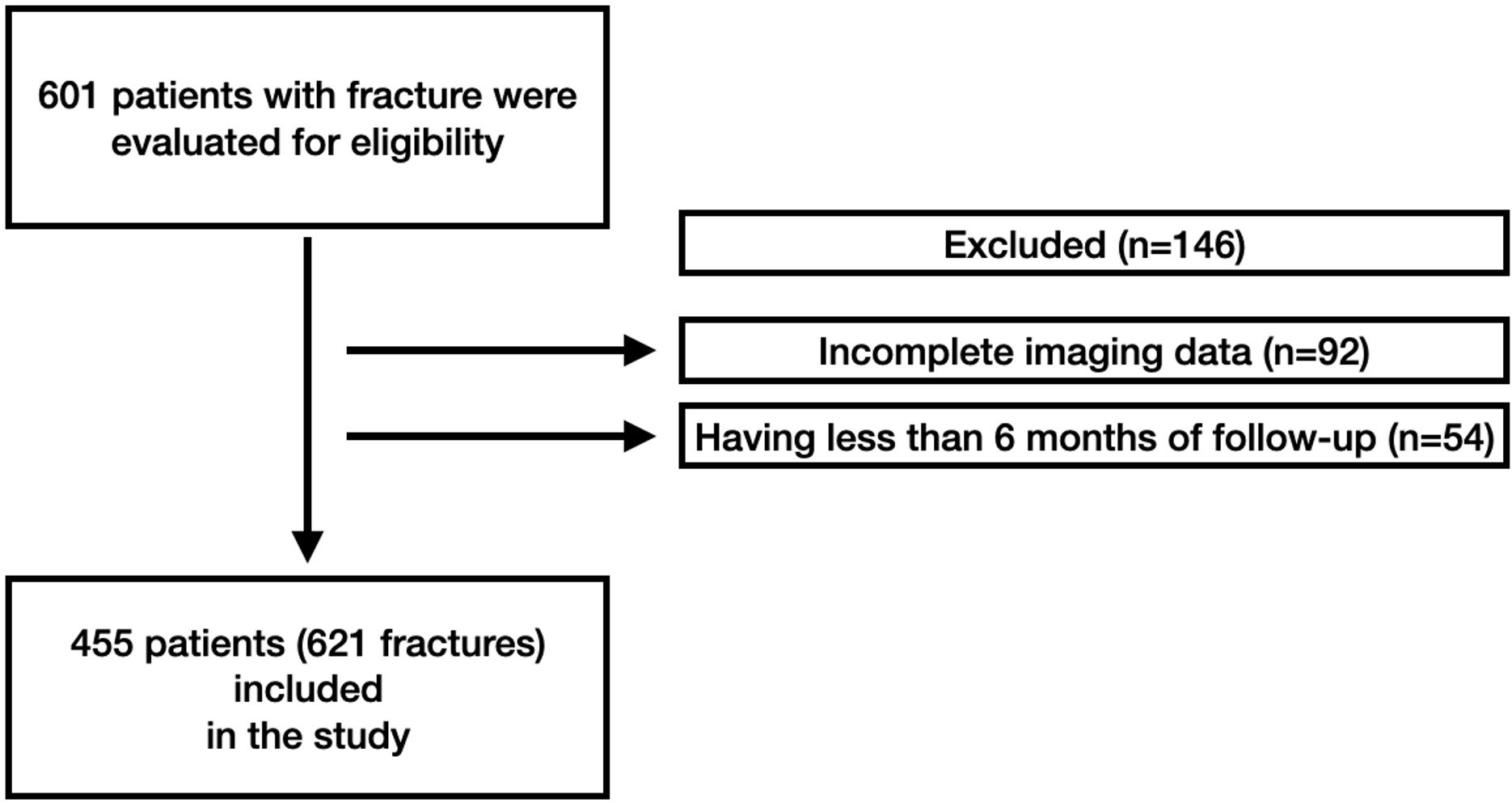
The study included 455 patients with 621 fractures Figure 1. The mean age for the study patients was 34.2 ± 24.3 (range; 6 – 93) years. 58% of the patients were male and 42% of the patients were female. The mean follow-up was 16 [range; 9 – 24] months. Associated medical comorbidities were noted in 34.5% of the patients. According to radiological findings, physical examinations and RUST scores the overall fracture union rate was 97,2% at 6th months follow-up. The RUST scores at the 6th-month follow-up was 10.5 which showed union of the fractures. The evaluation of RUST scrores during follow-ups were demonstrated in Figure 2. The overall complication rate was 7,3% following fracture fixation surgery.
The most common location for surgically treated fractures was ankle fractures (99 fractures) followed by forearm fractures (80 fractures) and hip fractures (52 fractures). The anatomic distribution of the fractures were demostrated in Table 2.
| Table 2. The distrubition of fracture types | |||
| Fracture types |
|
Fracture types |
|
| Proximal femur fracture |
|
Tibia plateau fracture |
|
| Clavicula fracture |
|
Trimalleolar fracture |
|
| Distal humerus fracture |
|
Proximal ulna fracture |
|
| Lateral malleolar + Bimalleolar fracture |
|
Femur Shaft fracture |
|
| Tibia shaft fracture |
|
Metatarsal fracture |
|
| Distal radius fracture |
|
Metacarpal fracture |
|
| Calcaneus fracture |
|
Ulna shaft fracture |
|
| Proximal humerus fracture |
|
Radial shaft fracture |
|
| Distal femur fracture |
|
Radial head fracture |
|
| Humerus shaft fracture |
|
Talus fracture |
|
| Distal tibia fracture |
|
Phalanx fracture |
|
| Both bone forearm fracture |
|
||
| TOTAL |
|
||
Postoperative complications were seen in 7.3% of the patients, while 92.7% of the patients were free of complications. In the postoperative course 6.2% of the patients suffered postoperative wound problems that were treated with antibiotics and surgical debridement when needed. Nonunion was seen in 1.3% [6] of the patients who were treated with revision surgery. Implant failure occurred in 1.3% [6] patients which caused revision fixation procedure. One patient with both bone forearm fracture suffered radial nerve palsy that was solved at the 3rd month follow up.
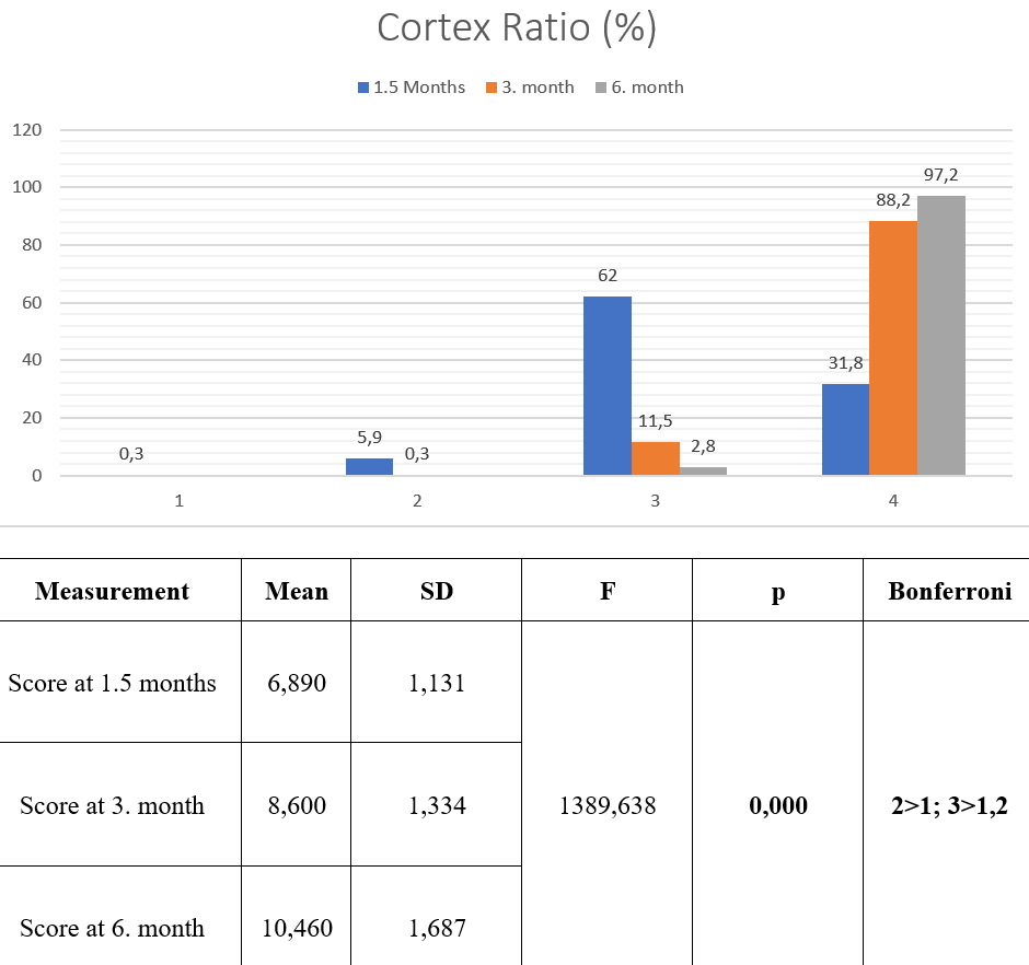
The fracture union rate was evaluated via RUST scores which significantly improved during follow-ups, Figure 3 . At the 6th weeks follow up, 62% of the fractures showed 3 and 31,8% showed 4 cortex callus formation. At 6th months follow up 97.2% of the fractures showed 4 cortices callus formation that improved significantly (p< 0.05).
A total of 996 different implants were used during the surgical treatment course. The distribution of the implants used during the surgical treatment were demonstrated in Table 3.
| Table 3. The distrubition of implants used in the study | |||
| Plates |
|
Plates |
|
| Mini Plate |
|
Olecranon Plate |
|
| 3.5 LC-LCP compression Plate |
|
LCP Superior Anterior Clavicle Plate, Locked Lateral Extension |
|
| 3.5mm Locking Reconstruction Plate |
|
Pediatric Plate 3.5 mm |
|
| Distal Radius Plate Anatomic |
|
LCP Proximal Medial Tibia Plate |
|
| 1/3 Tubular Plate |
|
Pediatric Plate 4.5 mm |
|
| Distal Lateral Femur Plate + Minimal Invasive |
|
4.5 mm Reconstruction Locking Plate |
|
| Distal Fibula 3.5mm Plate Anatomic |
|
T-Buttres Plate |
|
| Distal Medial Humerus Plate |
|
Distal Lateral Dorsal Humerus Plate |
|
| Distal Medial Tibia Plate |
|
LCP Clavicle Hook Plate |
|
| 4.5mm Compression Plate (LC-LCP) (Narrow + Broad ) |
|
T-Locking Plate |
|
| Proximal Humerus Plate Anatomic |
|
LCP Radius T-Plate, Bend angle |
|
| Proximal Femur Plate |
|
LCP Superior Anterior Clavicle Plate |
|
| Proximal Tibia Lateral Plate |
|
L-Buttress Plate |
|
| Distal Tibia Lateral Compression Plate |
|
Distal Ulna Hook Plate |
|
| Calcaneus Plate |
|
||
| Total (Plates) |
|
||
| Nails |
|
Cannulated screws |
|
| Cannulated Tibia Nail |
|
Cannulated screw 4.5mm |
|
| Titanium Elastic Nail |
|
Cannulated screw 3.5mm |
|
| Proximal Femoral Nail |
|
Headlless Compression Screw |
|
| Long Reconstruction Femoral Nail |
|
Cannulated screw 6.5mm |
|
| Humerus Nail |
|
Cannulated screw 7.3mm |
|
| Total (Nails) |
|
Total (Cannulared Screws) |
|
The most important aspect of orthopedic trauma implants is to bear biological loads without breakage till fracture union. Since the clinical outcomes depends on mutlifactorial viariables like fracture type, location, patient related ractors like comorbidities, treatment compatibility , and surgical factors like techniqe and proper implant usage, we decided to analyze implant survival till union and operative complications. Below are the individual analysis of specific implants.
Proximal humerus anatomic plate
The proximal humerus anatomic plate (Oltho medical) allows sending locking screws in various directions to improve stabilization and stabilize humeral head via rotator cuff sutures fixed on the plate through holes. 28 proximal humerus anatomic plates were used for the fixation of proximal humerus fractures. The union rate for the proximal humerus fractures was 100%. The implant survival rate for the proximal humerus plate until fracture union was 100%. No implant related complication was observed in study population.
Clavicle plates
Oltho medical clavicle plates include superior anterior locking compression clavicle plates and clavicle hook plates. Superior clavicle plate also has lateral extended version that allow sending locking 2.4mm screws to lateral fragments. A total of 34 clavicle plates were used in this study. The fracture union rates for clavicle shaft fractures was 100%. The implant survival rate for superior anterior locking compression clavicle plates till fracture union is 100%. No implant related complication was observed in the study population.
Oltho medical LCP clavicle hook plates allow treatment of distal clavicle fractures and acromioclavicular joint injuries. The healing rate for the distal clavicle fractures was 100%. The implant survival rate for the LCP clavicle hook plate till fracture union was 100%. No implant related complication was observed in study population. 10 patients received implant removal surgery at the 6 months follow up as routine elective implant removal.
Distal humerus plates
Oltho medical distal humerus plates included medial and lateral dorsal column plates used for fixation of distal humerus fractures. A total of 49 distal humerus medial and lateral dorsal plates were used for the fixation of distal humerus fractures. The union rate for the distal humerus fractures was 96%. There were no implant breakage in our medical records. Two fractures received revision fixation surgery with bone grafting, and both fractures healed uneventfully.
Olecranon plates
Oltho medical olecranon plates allow fixation olecranon fractures and olecranon osteotomies. 25 olecranon plates were analyzed. No implant breakage was recorded. The union rates for the olecranon fractures and olecranon ostetomies was 90%. Two patients received revision fixation surgery. Implant prominence and irritation was noted on 25% of the patients that is paralel to the literature.
Distal radius plates
Oltho medical distal radius plate included anatomic distal radius anatomic plates and LCP radius T-plate with bent angle. Anatomic distal radius plate are utilized for fixation of distal radius fractures. Locking distal screw configuration allows restoration of joint line and volar tilt. A total of 35 distal radius plates were analyzed. All fractures were healed uneventfully at the 3rd month follow up. No implant breakage was seen. Irritation of the extensor tendons was recorded due to the overextending screws in 3 patients (9%) which were removed following fracture union.
Proximal femur plates
Oltho medical proximal femur plates were used for the fixation of the proximal femur fractures, femoral osteotomies and trochanteric fractures. 28 proximal femur plates were analyzed. Union rate for the proximal femur fractures was 96%. One patient had trochanteric non-union which was asymptomatic and left untreated. No implant breakage was recorded.
Distal femur plates
Oltho medical distal femoral lateral plates were used for fixation of the distal femur fractures with or without cannulated screws. 26 distal femur plates were analyzed. The union rate for distal femur fractures were 100%. No implant breakage was observed.
Proximal tibia plates
Oltho medical proximal tibia plates included 4.5mm LCP medial plates and LCP lateral proximal tibia plates. A total of 48 proximal tibia plates were analyzed in 27 fractures. One patient suffered from non union that was successfully treated via revision fixation and bone grafting. Healing rate for the proximal tibia fractures was 96%. No implant breakage was noted.
Distal tibia plates
Oltho medical distal tibia plates included distal medial tibial plate and distal lateral compression plates. A total of 47 plates in 40 patients were analyzed. The union rate for distal tibia fractures were 92.5%. Three patients suffered from nonunion and received revision surgery. Screw breakage was seen in 2 (5%) patients due to nonunion, in 1 patient infection and soft tissue problems caused nonunion. No plate breakage was recorded.
Fibula plate
Thirty-two distal fibula anatomic plate was analyzed in 32 distal fibula fractures. The healing rate of the fibula fractures was 100%. No implant related problems were recorded. All of the implants survived throughout the treatment course.
Mini plates
Oltho medical Mini plates were used in various type of fracture fixation constructs like primary fixation providers in small bones as metatarsal and phalangeal fractures or supplementary fixation as anti rotation plates and neutralization plates. 36 mini plates, and a total of 37 L buttress, T buttress, and T locking plates were analyzed. There were no implant breakage recorded.
3.5 and 4.5 reconstruction plates
Oltho medical 3.5 and 4.5 reconstruction plates were used in primary or secondary fixation providers. A total of 53 reconstruction plates were analyzed and no implant failure was recorded in our study cohort.
3.5- 4.5 Compression plates [LC-DCP]
Oltho Medical 3.5 -4.5 compression plates provides compression of the fracture ends with cortical screws. Additionally adding locking screws to the construct would increase the rotational stability. A total of 65 LC-DCP plates were analyzed and no implant breakage was recorded.
Nails
A total of 130 intramedullary nails were analyzed in this study. Below are the specific data for the implants.
Femoral Nails: There was 1 nail breakage due to subtrochanteric fracture non-union and 1 interlocking screw breakage due to segmentary femur fracture out of 56 femoral nails that ended up with fracture union following implant removal and revision fixation surgery with the implant survival rates was 96.4% for Oltho medical femoral nails.
Tibia Nails: No nail breakage was recorded in 32 Oltho medical cannulated tibia nails. There were 2 interlocking screw breakages in 2 distal tibia fractures and 1 interlocking screw breakage tibia shaft breakage in an open tibia shaft fracture. Three cases needed exchange naill revision surgery all ended up with union.
Humerus nails: A total of 12 humerus intramedullary nails were analyzed and no implant breakage was observed in this study. All fractures healed uneventfully.
Titanium elastic nails: Oltho medical titanium elastic nails were utilized in pediatric trauma surgeries like tibia , femur and forearm fractures. Among 30 nails that were analyzed no implant breakage was recorded. All of the titanium elastic nails were removed following fracture union.
Cannulated screws and headless compression screws
Oltho medical cannulated screws and headless compression screws were used in the treatment of periarticular distal femur, proximal tibia, distal tibia, proximal femur, small bone fractures like scaphoid, metacarpals, phalangeal fractures foot fractures. Headless compression A total of 177 cannulated screws were analyzed and 1 implant breakage was recorded in a Lisfranc injury fixed with 4.0 headless cannulated screw. This implant breakage was attributed to early weight bearing.
Discussion
The present study demonstrated a high overall fracture union rate of 97.2% following surgical fixation, with implant failure observed in only 6 patients (1.3%). The overall implant survival rate of Oltho Medical trauma implants was 98.7%, indicating reliable biomechanical performance across a wide range of fracture types. Importantly, all fractures ultimately achieved union after appropriate management, underscoring the clinical effectiveness and safety of these implants in routine orthopedic trauma practice.
With every surgical procedure and implantation in orthopaedics there starts a race between implant failure and healing process of fracture [7]. Implant failure can be in form of plastic deformation, corrosion, or fatigue failure. Mechanical failure of implant can result due to repetitive loading and stress. In absence of union even the toughest metallic device and best designed implants are known to fail [13].
Technical aspects of implant failure have been studied in various studies [11,13] [22] [23]. According to laboratory results from two studies Azevedo in Brazil and Amel Farad H in Iran, manufacturers did not follow the standards in their cases of failed implant analysis. It is possible that implants differ in terms of purity and alloy inclusion from manufacturer to manufacturer [23]. In another study by Barbosa on three cases of implant failure, surgical technique and implant design, selection of implant have been reported to be important cause of implant failure [22]. Studies on implant quality control and designing errors are out of scope of this research and are limiting factors.
In study by Sharma et al. 2.4% implant failure was associated with deep infection[22]. In our study infection was found in 10 cases (2.2%). This infection was superficial to deep and associated with loosening of implant. Infection could have occurred during the surgery due to faulty surgical technique of open reduction and fixation of implant. Re-trauma is also major factor of implant failure during consolidation phase of fracture healing. Sharma et al. have also found re-trauma as significant cause for implant failure in their study.
Excessive body weight of the patient and early weight bearing on affected lower limb imparts more stress on implant during the healing stage of fracture. During the stance phase of gait cycle, load on lower limb is more than three times the body weight. Alfred O. Ogbemudia et al. in their study found patient non compliance and excessive body weight as significant reason for failure of implant and suggested cautious ambulation and graduated weight bearing [24]. Similar observations have been reported by Sharma et al., who emphasized that premature loading before sufficient callus formation increases the risk of screw loosening and fatigue failure [22]. Biomechanical analyses also demonstrate that repetitive cyclic loading under excessive weight significantly accelerates microcrack propagation within the implant material, particularly in stainless steel constructs [13,25]. In the present study, all instances of implant breakage occurred in patients with delayed union or early weight bearing, supporting these findings. Therefore, appropriate patient education, gradual rehabilitation, and close radiographic follow-up are essential to minimize mechanical overload and optimize implant longevity.
Plastic deformation, brittle and fatigue failure are known to occur with minor loads and re-trauma. 1.5% fatigue failure was observed in our study. Fatigue failure is associated with poor design, workman ship, handling and implant breaks from cyclical loading [7]. Surface notches or holes severely affect fatigue strength. Scratches or corrosion can also reduce the strength and predispose to implant failure. Fatigue failure of plates is more common than nails as intramedullary location of nails in shaft prevents some bending forces responsible for fatigue failure [13]. Plate ends act as stress riser leading to fresh fractures at ends. Plate fixation requires perfect reduction and anatomical reconstruction and may interfere with periosteal blood supply[26]. Poorly fixed implant with excessive soft tissue handling leads to failure of union and implant[26]. Recent biomechanical studies have demonstrated that even minor surface imperfections, such as micro-notches or corrosion pits, can initiate fatigue cracks and markedly reduce implant lifespan under repetitive cyclic loading [27]. Furthermore, comparative studies have shown that while plate constructs provide higher initial stiffness, intramedullary nails display superior fatigue endurance due to their load-sharing nature and reduced exposure to bending stresses [28]. These findings are in accordance with our observations, suggesting that implant longevity depends not only on material quality but also on mechanical design and surgical technique.
Intra-medullary implants are load sharing and provide good stability to fractures of long bone, allowing early rehabilitation and functional recovery of patient. Locked intra-medullary nail provide excellent axial and rotational stability as compared to unlocked intra-medullary nail. Intra-medullary implant failures occur with small diameter nail, improper selection of implant which is not suitable for unstable fracture configurations. Failure to provide rotational stability at fracture site leads to loosening of implant and failure [11]. At times inability to select a suitable implant to match the fracture configuration or an improper surgical technique to restore fracture anatomy, inadequate fixations, pre/ post-operative complications like infections, patient non-compliance with implant instructions and degree of union lead to failure of implant [25].
We identified 1 cephalomedullary nail breakage in a subtrochanteric femur fracture 12 months after the surgery caused by nonunion of the fracture. The nail breakage occurred in the shaft-neck intersection and this type of failure is typically seen because of metal fatigue due to excessive loading following fracture non-union. In 2 patients with distal tibia fracture, broken screws of the distal medial tibia plate was observed. This was attributed to early weight bearing on the extremity and extended fracture union time. Broken interlocking screws of the femoral nail was noted 1 one, tibial nail in 1 patient. Interlocking screws of the femoral and tibia nails occured following delayed union and cause auto-dynamization of the fracture. Both fractures healed uneventfully. Additionally, 1 4mm headless compression screw was broken following surgical treatment of Lisfranc injury at the 6 months follow-up. The screw breakage was attributed to the motion between fixed fragments due to weight bearing. No implant removal was required.
The major limitation of this study was its retrospective nature. Prospective randomized studies would be the ideal way of determining implant success in trauma surgery. However, fracture healing rates with a minimum of 6 months follow-up would provide sufficient information for survival analysis. Additionally, this study lacks clinical outcomes. In trauma surgery, clinical outcomes are more related to the fracture type, injury pattern, patient specific variables like age, associated comorbidities and activity levels. Thus, analyzing clinical outcomes were beyond the scope of this study.
Conclusion
In conclusion medical trauma implants provided good to excellent survivability compared to the literature till fracture healing. Even though complications were noticed during follow-ups all were successfully managed with appropriate approach.
Ethical approval
This study was approved by the Hacettepe University Health Sciences Research Ethics Committee (Date: 02.05.2022, Decision/Protocol No: IRB: GO22/942). Informed consent was obtained from all participants involved in this study.
Data availability statement
The data that support the findings of this study are available from the corresponding author upon reasonable request.
Conflict of interest
The authors declare that this study was conducted in the absence of any commercial or financial relationships that could be construed as a potential conflict of interest.
Funding
The authors declare that this study received no funding.
Generative AI statement
The authors declare that no generative AI or AI-assisted technologies were used in the writing or preparation of this study.
References
- Einhorn TA, Gerstenfeld LC. Fracture healing: mechanisms and interventions. Nat Rev Rheumatol 2015;11(1):45-54. https://doi.org/10.1038/nrrheum.2014.164
- Phillips AM. Overview of the fracture healing cascade. Injury 2005;36(Suppl 3):S5-7. https://doi.org/10.1016/j.injury.2005.07.027
- Marsell R, Einhorn TA. The biology of fracture healing. Injury 2011;42(6):551-5. https://doi.org/10.1016/j.injury.2011.03.031
- Bishop JA, Palanca AA, Bellino MJ, Lowenberg DW. Assessment of compromised fracture healing. J Am Acad Orthop Surg 2012;20(5):273-82. https://doi.org/10.5435/JAAOS-20-05-273
- Giannoudis PV, Jones E, Einhorn TA. Fracture healing and bone repair. Injury 2011;42(6):549-50. https://doi.org/10.1016/j.injury.2011.03.037
- Rath B, Nam J, Knobloch TJ, Lannutti JJ, Agarwal S. Compressive forces induce osteogenic gene expression in calvarial osteoblasts. J Biomech 2008;41(5):1095-103. https://doi.org/10.1016/j.jbiomech.2007.11.024
- Carter DR, Beaupré GS, Giori NJ, Helms JA. Mechanobiology of skeletal regeneration. Clin Orthop Relat Res 1998;355:S41-55. https://doi.org/10.1097/00003086-199810001-00006
- Shapiro F. Cortical bone repair. The relationship of the lacunar-canalicular system and intercellular gap junctions to the repair process. J Bone Joint Surg Am 1988;70(7):1067-81.
- Gerstenfeld LC, Alkhiary YM, Krall EA, et al. Three-dimensional reconstruction of fracture callus morphogenesis. J Histochem Cytochem 2006;54(11):1215-28. https://doi.org/10.1369/jhc.6A6959.2006
- Perren SM. Evolution of the internal fixation of long bone fractures. The scientific basis of biological internal fixation: choosing a new balance between stability and biology. J Bone Joint Surg Br 2002;84(8):1093-110. https://doi.org/10.1302/0301-620x.84b8.13752
- Kuzyk PR, Schemitsch EH. The basic science of peri-implant bone healing. Indian J Orthop 2011;45(2):108-15. https://doi.org/10.4103/0019-5413.77129
- Shaikh M, Kahwash F, Lu Z, Alkhreisat M, Mohammad A, Shyha I. Revolutionising orthopaedic implants-a comprehensive review on metal 3D printing with materials, design strategies, manufacturing technologies, and post-process machining advancements. The International Journal of Advanced Manufacturing Technology 2024;134(3):1043-76. https://doi.org/10.1007/s00170-024-14218-y
- Kim T, See CW, Li X, Zhu D. Orthopedic implants and devices for bone fractures and defects: past, present and perspective. Engineered Regeneration 2020;1:6-18. https://doi.org/10.1016/j.engreg.2020.05.003
- Li Z, Kuhn G, von Salis-Soglio M, et al. In vivo monitoring of bone architecture and remodeling after implant insertion: the different responses of cortical and trabecular bone. Bone 2015;81:468-477. https://doi.org/10.1016/j.bone.2015.08.017
- Zura R, Xiong Z, Einhorn T, et al. Epidemiology of fracture nonunion in 18 human bones. JAMA Surg 2016;151(11):e162775. https://doi.org/10.1001/jamasurg.2016.2775
- Hammer RR, Hammerby S, Lindholm B. Accuracy of radiologic assessment of tibial shaft fracture union in humans. Clin Orthop Relat Res 1985;(199):233-8.
- Nicholson JA, Yapp LZ, Keating JF, Simpson AHRW. Monitoring of fracture healing. Update on current and future imaging modalities to predict union. Injury 2021;52(Suppl 2):S29-34. https://doi.org/10.1016/j.injury.2020.08.016
- Schwarzenberg P, Darwiche S, Yoon RS, Dailey HL. Imaging modalities to assess fracture healing. Curr Osteoporos Rep 2020;18(3):169-179. https://doi.org/10.1007/s11914-020-00584-5
- Whelan DB, Bhandari M, McKee MD, et al. Interobserver and intraobserver variation in the assessment of the healing of tibial fractures after intramedullary fixation. J Bone Joint Surg Br 2002;84(1):15-8. https://doi.org/10.1302/0301-620x.84b1.11347
- Leow JM, Clement ND, Tawonsawatruk T, Simpson CJ, Simpson AHRW. The radiographic union scale in tibial (RUST) fractures: reliability of the outcome measure at an independent centre. Bone Joint Res 2016;5(4):116-21. https://doi.org/10.1302/2046-3758.54.2000628
- Whelan DB, Bhandari M, Stephen D, et al. Development of the radiographic union score for tibial fractures for the assessment of tibial fracture healing after intramedullary fixation. J Trauma 2010;68(3):629-32. https://doi.org/10.1097/TA.0b013e3181a7c16d
- Sharma AK, Kumar A, Joshi GR, John JT. Retrospective study of implant failure in orthopaedic surgery. Med J Armed Forces India 2006;62(1):70-2. https://doi.org/10.1016/S0377-1237(06)80164-4
- Peivandi MT, Yusof-Sani MR, Amel-Farzad H. Exploring the reasons for orthopedic implant failure in traumatic fractures of the lower limb. Arch Iran Med 2013;16(8):478-82.
- Ogbemudia AO, Umebese PFA. Implant failure in osteosynthesis of fractures of long bones. Journal of Medicine and Biomedical Research 2009;5(2). https://doi.org/10.4314/jmbr.v5i2.10696
- Chen CE, Weng LH, Ko JY, Wang CJ. Management of nonunion associated with broken intramedullary nail of the femur. Orthopedics 2008;31(1):78. https://doi.org/10.3928/01477447-20080101-07
- Beeharry MW, Ahmad B. Principles of fracture healing and fixation: a literature review. Cureus 2024;16(12):e76250. https://doi.org/10.7759/cureus.76250
- Wu K, Li B, Guo JJ. Fatigue crack growth and fracture of internal fixation materials in in vivo environments-a review. Materials (Basel) 2021;14(1):176. https://doi.org/10.3390/ma14010176
- Kandemir U, Herfat S, Herzog M, Viscogliosi P, Pekmezci M. Fatigue failure in extra-articular proximal tibia fractures: locking intramedullary nail versus double locking plates-a biomechanical study. J Orthop Trauma 2017;31(2):e49-54. https://doi.org/10.1097/BOT.0000000000000729
Copyright and License
Copyright © 2026 The Author(s). This is an open access article distributed under the Creative Commons Attribution License (CC BY), which permits unrestricted use, distribution, and reproduction in any medium or format, provided the original work is properly cited.