

Abstract
Objective: The coexistence of primary hyperparathyroidism (PHPT) and papillary thyroid carci-noma (PTC) has been increasingly recognized; however, its impact on tumor behavior and clinicopathological features remains unclear. To investigate the association be-tween PHPT and the clinicopathological characteristics of PTC in a large single-center cohort.
Materials and Methods: This retrospective study included patients who underwent parathyroidectomy for PHPT between 2019 and 2024. Patients with concomitant PTC were identified and compared with a separate cohort of patients with PTC without PHPT. Demographic, biochemical, and clinicopathological features, including tumor subtypes and adverse pathological characteristics, were analyzed.
Results: Among 190 patients with PTC, 91 (47.9%) had concomitant PHPT. Patients with PHPT were older and more frequently female. Tumor size was significantly smaller in patients with PHPT. Aggressive histological subtypes were significantly less frequent in the PHPT group (8.8% vs. 21.2%, p = 0.017). In addition, capsular invasion, lym-phovascular invasion, perineural invasion, and lymph node metastasis were observed less frequently in patients with PHPT. Radioactive iodine use was also significantly lower in this group.
In the PHPT cohort (n = 750), the presence of concomitant PTC was not associated with significant differences in preoperative calcium, phosphorus, PTH, or ALP levels. However, patients with PTC had lower preoperative magnesium levels and exhibited distinct postoperative biochemical profiles.
Conclusion: PHPT may be associated with non-aggressive subtypes and more favorable clinico-pathological features in PTC. However, these findings should be interpreted cautious-ly, as tumor size and other potential confounders may influence the observed associa-tions. PHPT alone should not be considered a determinant for treatment de-escalation, and clinical decision-making should remain guided by established risk-adapted strategies. These findings may provide additional insight into risk stratification in patients with coexisting PHPT and PTC.
Keywords: hyperparathyroidism, thyroid cancer, papillary
Introduction
Primary hyperparathyroidism (PHPT) is a common endocrine disorder affecting ap-proximately 0.1–0.4% of the general population [1]. In the majority of cases, it is caused by a solitary parathyroid adenoma, and surgical excision remains the definitive treatment [2]. Given the relatively high prevalence of con-comitant thyroid pathology, preoperative thyroid evaluation is routinely recommended in patients undergoing parathyroidectomy [3].
Papillary thyroid carcinoma (PTC), accounting for 85–90% of all thyroid malignancies, is the most common type of thyroid cancer, with a steadily increasing incidence worldwide over recent decades [4,5]. Although several stud-ies have reported an association between PHPT and thyroid carcinoma [6,7], the nature of this relationship remains incompletely understood [8]. In particular, it is still unclear whether this coexistence reflects a causal link or represents a coincidental finding, and further investigation is required to clarify the underlying mechanisms.
Potential shared molecular pathways and genetic predispositions contributing to the coexistence of these two conditions have been proposed, but current evidence re-mains limited [9,10]. Moreover, comprehensive studies eval-uating the impact of PHPT on the prognosis, disease progression, and treatment out-comes of PTC—as well as the potential influence of PTC on the clinical course of PHPT—are scarce [11,12].
A better understanding of the clinical implications of this coexistence is of considera-ble importance, particularly in terms of diagnostic challenges, surgical planning, and treatment strategies [7,12-14].
Therefore, the aim of this study was to systematically evaluate the potential interac-tion between PHPT and PTC in a large patient cohort.
Materials and Methods
Study design and patient selection
This retrospective study was conducted in two phases. In the first phase, all consecu-tive patients with PHPT who underwent surgery at our institution between January 2019 and December 2024 were included. All procedures were performed by attending surgeons from the Department of General Surgery.
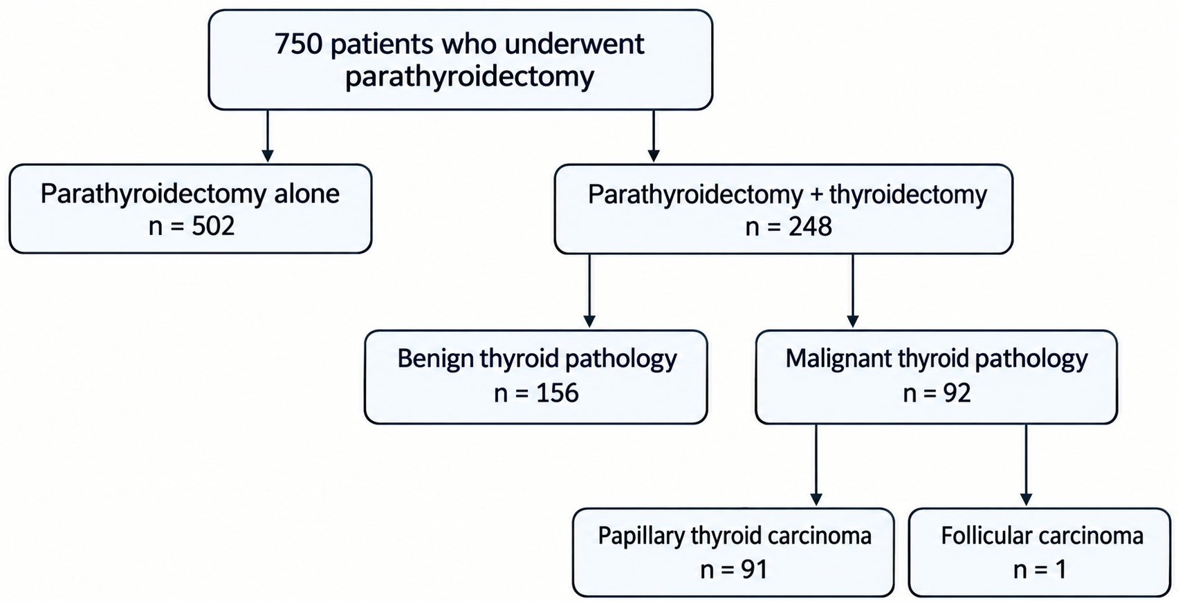
During this period, a total of 816 patients underwent parathyroidectomy. Of these, 67 patients were excluded due to secondary or tertiary hyperparathyroidism (n = 36), fa-milial syndromes such as MEN1 or MEN2A (n = 7), incomplete medical records (n = 17), or unsuccessful parathyroidectomy (n = 6). After applying these exclusion criteria, 750 patients were included in the final analysis and were stratified according to the presence or absence of concomitant PTC (Figure 1).
In the second phase, to enable comparison between patients with PTC with and with-out PHPT, a separate cohort of patients with PTC without PHPT was included. These patients were selected from consecutive cases who underwent surgery between Jan-uary 2019 and December 2020 and had a histopathological diagnosis of PTC, in order to achieve a comparable sample size with the PTC + PHPT group. This approach was chosen to minimize temporal bias and ensure a homogeneous surgical and diagnostic environment following the establishment of our high-volume center in 2019.
Patients with concomitant PHPT and PTC were included in both analytic cohorts, as each analysis addressed a distinct research question.
Data collection
Preoperative and postoperative biochemical data were retrieved from institutional electronic medical records. All biochemical analyses were performed in the central laboratory of our institution using standardized automated analyzers in accordance with the manufacturers’ instructions.
Alkaline phosphatase (ALP), calcium, phosphorus, magnesium, and creatinine levels were measured using the Siemens Atellica CH automated chemistry analyzer (Sie-mens Healthineers, Erlangen, Germany), employing IFCC-recommended kinetic col-orimetric or enzymatic methods, as appropriate. Parathyroid hormone (PTH) levels were measured using chemiluminescent immunoassay on the Siemens Atellica IM analyzer.
The reference ranges were as follows: ALP 42–98 U/L, calcium 8.7–10.4 mg/dL, phosphorus 2.4–5.1 mg/dL, magnesium 1.3–2.7 mg/dL, and creatinine 0.5–1.1 mg/dL. The reference range for serum PTH was 18.4–80.1 ng/mL.
Serum calcium concentrations were corrected for albumin using the following formula:
Corrected calcium (mg/dL) = [0.08 × (40 − albumin (g/L))] + measured calcium.
Laboratory parameters were collected at predefined perioperative time points. Magne-sium and phosphorus levels obtained during the final week prior to surgery were rec-orded. PTH levels included the highest preoperative value measured within one month before surgery and the postoperative value measured on postoperative day 1. Similar-ly, calcium levels were defined as the highest preoperative value within one month prior to surgery and the postoperative value measured on postoperative day 1.
Clinical and pathological data
Data on PTC were obtained from pathology reports. Parathyroid adenoma localization and dimensions were determined based on preoperative ultrasonography and opera-tive records. Adenomas were assumed to have an ellipsoid shape, and their volumes were calculated using the formula: volume = (π/6) × length × width × depth.
Patients with renal stones detected on imaging (ultrasonography or computed tomog-raphy) or with a documented history of nephrolithiasis were classified as having a his-tory of renal stones.
Definitions
PHPT with PTC was defined as the coexistence of histopathologically confirmed para-thyroid tumor and PTC in the same patient, with both lesions identified and treated during the same surgical procedure.
PTC subtypes were classified according to the World Health Organization criteria.
Aggressive PTC subtypes were defined as tall cell, hobnail, columnar cell, diffuse sclerosing, and solid/trabecular variants, based on their established association with adverse clinicopathological features. Other subtypes—including classic (conventional), follicular, Warthin-like, oncocytic, and cribriform–morular variants—were not classified as aggressive.
Bone health was assessed using bone mineral density (BMD) and T-scores obtained from dual-energy X-ray absorptiometry (DXA) of the lumbar spine. Lumbar spine measurements were selected because they represented the most consistently availa-ble data in this retrospective cohort (missing in only 8 patients). Measurements from the radius, femoral neck, and total hip were not consistently available and were there-fore excluded from analysis.
DXA measurements were performed using a GE/Lunar Prodigy densitometer (GE Healthcare, Madison, WI, USA) according to standard protocols. The precision error for lumbar spine BMD measurements was within acceptable limits, with a coefficient of variation (CV) ≤ 2%, in accordance with international densitometry standards.
Statistical analysis
Continuous variables were assessed for normality using visual inspection and the Shapiro–Wilk test and are presented as mean ± standard deviation (SD) or median with interquartile range (IQR), as appropriate. Categorical variables are expressed as frequencies and percentages.
Comparisons between groups were performed using Student’s t-test or the Mann–Whitney U test for continuous variables and the chi-square test or Fisher’s exact test for categorical variables, as appropriate.
A two-sided p-value < 0.05 was considered statistically significant. All statistical anal-yses were performed using R software (version 4.1.1).
Ethical approval
This study was conducted in accordance with the ethical principles of the Declaration of Helsinki. The study protocol was approved by the local Ethics Committee (approval number: TABED 1-25-1568). Informed consent was obtained from all participants as part of the routine institutional admission procedure, which includes permission for the retrospective use of anonymized clinical data for scientific research purposes.
Results
Comparison of PTC with and without concomitant PHPT
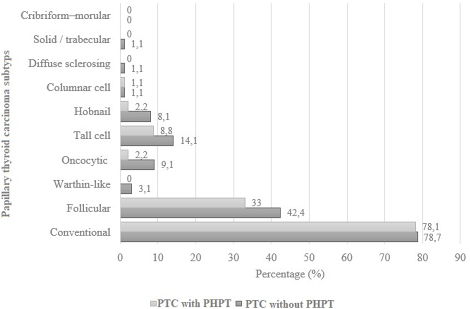
Among 190 patients with PTC, 91 (47.9%) had concomitant PHPT. Patients with PTC and PHPT were older than those without PHPT (55 ± 11 vs. 47 ± 14 years, p < 0.001) and were more frequently female (89% vs. 71.7%, p = 0.003). The distribution of con-ventional (classic) PTC was similar in patients with and without PHPT (78.1% vs. 78.7%). In contrast, the follicular subtype was more common in patients without PHPT (42.4% vs. 33.0%) (Figure 2). Aggressive PTC subtypes were more frequently ob-served in patients without PHPT (21.2% vs. 8.8%, p = 0.017).
Patients without PHPT demonstrated higher rates of adverse pathologic features, in-cluding capsule invasion (31.3% vs. 17.6%, p = 0.028), lymphovascular invasion (20.2% vs. 6.6%, p = 0.006), perineural invasion (11.1% vs. 3.3%, p = 0.039), and central lymph node metastasis (29.3% vs. 6.6%, p < 0.001). Lateral lymph node me-tastasis was observed exclusively in patients without parathyroid adenoma (PA) (7.9% vs. 0%, p < 0.001). Radioactive iodine therapy was administered more frequently to patients without PHPT (80.8% vs. 21.1%, p < 0.001). The rates of multifocality, bilat-erality, recurrence, and follow-up duration did not differ significantly between the groups (Table 1).
| IQR: Interquartile range, PHPT: Primary Hyperparathyroidism, PTC: Thyroid Papillary Carcinoma, SD: Standard Deviation. | ||||
| Table 1. Comparison of PTC with or without PHPT | ||||
| Variables |
|
|
|
|
| Age, years, mean, SD |
|
|
|
|
| Sex, female, n, % |
|
|
|
|
| Tumor Size, cm, median, IQR |
|
|
|
|
| Aggressive tumor variant, n, % |
|
|
|
|
| Multifocal tumor, n, % |
|
|
|
|
| Bilateral tumor, n, % |
|
|
|
|
| Capsule Invasion, n, % |
|
|
|
|
| Extrathyroidal extension, n, % |
|
|
|
|
| Lymphovascular Invasion, n, % |
|
|
|
|
| Perineural Invasion, n, % |
|
|
|
|
| Central Lymph Node Metastasis, n, % |
|
|
|
|
| Lateral Lymph Node Metastasis, n, % |
|
|
|
|
| Radioactive Iodine Treatment, n, % |
|
|
|
|
| Recurrence, n, % |
|
|
|
|
| Follow op time, months, median, IQR |
|
|
|
|
Comparison of PHPT with and without concomitant PTC
A total of 750 patients with parathyroid pathology were included, of whom 91 (12.1%) had concomitant PTC. Patients with PHPT and concomitant PTC were slightly older than those without PTC (55 ± 11 vs. 52 ± 12 years, p = 0.002) and were more fre-quently female (89% vs. 80%, p = 0.03).
The preoperative biochemical parameters, including serum calcium, phosphorus, par-athyroid hormone (PTH) and ALP levels, were largely comparable between the groups. However, patients with concomitant PTC had significantly lower preoperative magnesium levels (median 1.99 vs. 2.07 mg/dL, p = 0.003).
Postoperatively, patients with PA and PTC had significantly lower calcium levels (me-dian 8.48 vs. 8.90 mg/dL, p < 0.001), higher phosphorus levels (3.50 ± 0.81 vs. 3.21 ± 0.64 mg/dL, p = 0.001), and lower PTH levels (median 10 vs. 17 pg/mL, p = 0.002). Parathyroid gland location, pathology subtype, size, and volume did not differ signifi-cantly between the groups. Similarly, the prevalence of renal stones, bone mineral density, and T-scores were comparable between patients with and without concomi-tant PTC (Table 2).
| ALP: Alkaline Phosphatase, BMD: Bone Mass Density, IQR: Interquartile range, PHPT: Primary Hyperparathyroidism, PTH: Parathyroid Hormone, PTC: Thyroid Papillary Carcinoma, SD: Standard Deviation. | ||||
| Table 2. Comparison of PHPT with or without PTC | ||||
| Variables |
|
|
|
|
| Age, years, mean, SD |
|
|
|
|
| Sex, female, n, % |
|
|
|
|
| Preoperative Laboratory Values | ||||
| ALP, U/L, median, IQR |
|
|
|
|
| Calcium, mg/dL, median, IQR |
|
|
|
|
| Phosphorus, mg/dL, mean, SD |
|
|
|
|
| Magnesium, mg/dL, median IQR |
|
|
|
|
| Creatinine, mg/dL, median, IQR |
|
|
|
|
| PTH, pg/mL, median, IQR |
|
|
|
|
| Postoperative Laboratory Findings | ||||
| Calcium, mg/dL, median, IQR |
|
|
|
|
| Phosphorus, mg/dL, mean, SD |
|
|
|
|
| PTH, pg/mL, median, IQR |
|
|
|
|
| Parathyroid location, n, % |
|
|||
| Left Superior |
|
|
|
|
| Left Inferior |
|
|
|
|
| Right Superior |
|
|
|
|
| Right Inferior |
|
|
|
|
| Intrathyroidal |
|
|
|
|
| Atypic |
|
|
|
|
| Parathyroid pathology n, % |
|
|||
| Adenoma |
|
|
|
|
| Atypic adenoma |
|
|
|
|
| Hyperplasia |
|
|
|
|
| Cancer |
|
|
|
|
| Parathyroid size, mm, median, IQR |
|
|
|
|
| Parathyroid volume mm3, median, IQR |
|
|
|
|
| History of Renal Stone, n, % |
|
|
|
|
| BMD g/cm3, median, IQR |
|
|
|
|
| T Score, median, IQR |
|
|
|
|
Discussion
In this large cohort study, we compared patients with PTC with and without concomi-tant PHPT in terms of demographic, biochemical, and clinicopathological characteris-tics. Our findings suggest that the presence of PHPT may be associated with differ-ences in tumor behavior and could represent a clinically relevant factor in the evalua-tion of PTC. In addition, we performed a secondary analysis comparing patients with isolated PHPT and those with concomitant PTC across similar parameters.
Previous studies investigating the coexistence of PHPT and PTC have predominantly focused on the prevalence of thyroid disease in patients with PHPT, frequently report-ing a high incidence of papillary thyroid microcarcinomas (PTMCs) and incidentally detected tumors in this population [14-17]. However, to the best of our knowledge, large-scale studies systematically comparing the distribution of aggressive histological subtypes and other adverse clinicopathological features be-tween PTC patients with and without PHPT remain limited. In the present study, we evaluated not only tumor frequency but also a wide range of prognostic parameters reflecting tumor biology and clinical behavior. Our findings suggest that PTC associat-ed with PHPT may exhibit a more indolent clinicopathological phenotype compared with isolated PTC. Nevertheless, a causal relationship cannot be established based on the current data.
The existing literature on this topic is heterogeneous. Beebeejaun et al. proposed that elevated PTH levels, hypercalcemia, and low 1,25-dihydroxyvitamin D levels may in-fluence the tumor microenvironment and thereby alter the biological behavior of PTC [8]. Similarly, Jeong et al. and Çetin et al. reported that PTC associated with PHPT may present with more aggressive histopathological and clinical features [6,7]. In contrast, Hu et al. found no significant difference in tumor aggressiveness between PTC patients with and without PHPT, while Tsai et al. suggested that PTC associated with PHPT may follow an indolent course, potentially due to earlier detection [18]. These conflicting findings highlight the need for further well-designed studies to clarify the nature of this association.
One of the most notable findings of our study is the significantly lower frequency of aggressive histological subtypes—such as tall cell and hobnail variants, which are well known to be associated with poor prognosis and higher invasive potential—in patients with concomitant PHPT [19]. In addition, rates of capsular invasion, lym-phovascular invasion, and perineural invasion were significantly lower in this group, while extrathyroidal extension showed a trend toward lower frequency. The lower rate of lymph node metastasis further supports the notion that these tumors may exhibit a more indolent biological behavior [20-22].
However, the significantly smaller tumor size observed in patients with concomitant PHPT should be considered an important confounding factor that may partially ac-count for the lower rates of invasion and metastasis. Therefore, it would be inappro-priate to attribute the less aggressive clinicopathological features solely to the pres-ence of PHPT without accounting for tumor size differences. In this context, tumor size should be taken into consideration when interpreting the observed differences between groups.Adjuvant treatment patterns may also reflect these differences in tu-mor characteristics. The lower rate of radioactive iodine (RAI) use in patients with concomitant PHPT may suggest that these tumors are more frequently classified into lower-risk categories. Given that current guidelines recommend a more selective use of RAI in low-risk PTC patients [23,24], this finding may indi-cate that unnecessary adjuvant treatments could potentially be avoided in this sub-group. However, the difference in RAI use should be interpreted cautiously, as it likely reflects risk-adapted clinical decision-making rather than intrinsic tumor biology.
It has been suggested that PTC associated with PHPT may be detected at smaller sizes due to increased diagnostic scrutiny, leading to a more indolent clinical course [25]. This raises the possibility that detection bias may contribute to the observed differences in tumor behavior. However, the lower frequency of aggressive histological variants observed in our study suggests that the indolent phenotype can-not be explained solely by earlier detection and may also reflect underlying biological differences.
From a biological perspective, the chronic hypercalcemic microenvironment character-istic of PHPT may modulate calcium-sensing receptor (CaSR)-mediated signaling pathways, thereby influencing cellular proliferation and differentiation processes [26,27]. In addition, the observed indolent phenotype may speculatively reflect differences in the underlying molecular landscape, including a lower prevalence of alterations associated with aggressive behavior, such as BRAFV600E and TERT promoter mutations [28]. However, these hy-potheses require validation in future molecular and prospective studies.
The lower frequency of aggressive histological variants in PTC associated with PHPT may also be considered in the context of the risk-adapted approach emphasized in the American Thyroid Association (ATA) 2025 guidelines. Current recommendations de-fine recurrence risk based on a combination of pathological features, with the accumu-lation of adverse characteristics leading to higher risk categories [24]. In this regard, the lower rates of aggressive subtypes, lymph node metastasis, and other invasive features observed in patients with PHPT suggest that these tumors may be more likely to fall into lower-risk categories. However, given the differences in tumor size between groups and the absence of multivariable analyses, caution is warranted when translating these findings into clinical decision-making.
In our secondary analysis comparing patients with PHPT with and without concomi-tant PTC, preoperative biochemical parameters were largely similar between groups. The absence of significant differences in serum calcium, phosphorus, PTH, and ALP levels suggests that the presence of PTC is not associated with the biochemical se-verity of PHPT. This finding supports the notion that the two conditions may largely follow independent biochemical courses. Interestingly, preoperative magnesium levels were significantly lower in patients with concomitant PTC. Given the potential role of magnesium in cellular proliferation and oxidative stress, this finding may indicate a possible interaction between mineral metabolism and thyroid tumor biology. Our re-sults are consistent with previous studies reporting an association between low serum magnesium levels and thyroid cancer [29,30]. Postoperative-ly, patients with concomitant PTC exhibited lower calcium and PTH levels and higher phosphorus levels, suggesting a different balance of mineral metabolism following surgery. However, the absence of differences in parathyroid gland size, volume, local-ization, and pathological characteristics suggests that these findings may be more closely related to surgical extent or perioperative factors rather than intrinsic disease burden. Furthermore, the similar rates of renal stone history, bone mineral density, and T-scores between groups indicate that end-organ involvement in PHPT appears to be independent of the presence of concomitant PTC. Taken together, these find-ings suggest that the coexistence of PTC does not substantially alter the systemic clinical manifestations of PHPT.
Several limitations of this study should be acknowledged. First, the retrospec-tive design and baseline differences between groups—particularly in age, sex, and tumor size—limit the ability to fully account for potential confounding factors. Second, the absence of multivariable analyses precludes definitive assessment of the inde-pendent effect of PHPT. Finally, the relatively short follow-up period restricts the eval-uation of recurrence and long-term oncological outcomes.
In conclusion, this study suggests that PHPT may be associated with non-aggressive subtypes of PTC and that these tumors may exhibit less aggressive clinicopathologi-cal features compared with isolated PTC. The lower frequency of aggressive histolog-ical variants, invasive features, and lymph node metastasis supports the notion of a more indolent tumor phenotype. While the more favorable pathological profile ob-served in patients with concomitant PHPT appears to be consistent with the risk-adapted approach emphasized in the ATA 2025 guidelines, PHPT itself should not be considered an independent criterion for treatment de-escalation. Clinical decision-making in this patient population should remain guided by established guidelines and multidisciplinary evaluation. These findings may provide additional insight into risk stratification in patients with coexisting PHPT and PTC.
Ethical approval
This study was approved by the Ankara Bilkent City Hospital-Medical Research and Clinical Ethics Evaluation Committee (Date: 13.08.2025, Decision/Protocol No: TABED 1-25-1568). Informed consent was obtained from all participants involved in this study.
Data availability statement
The data that support the findings of this study are available from the corresponding author upon reasonable request.
Conflict of interest
The authors declare that this study was conducted in the absence of any commercial or financial relationships that could be construed as a potential conflict of interest.
Funding
The authors declare that this study received no funding.
Generative AI statement
The authors declare that no generative AI or AI-assisted technologies were used in the writing or preparation of this study.
References
- Heath DA. Primary hyperparathyroidism. Clinical presentation and factors influencing clinical management. Endocrinol Metab Clin North Am 1989;18(3):631-46.
- Fraser WD. Hyperparathyroidism. Lancet 2009;374(9684):145-58. https://doi.org/10.1016/S0140-6736(09)60507-9
- Wilhelm SM, Wang TS, Ruan DT, et al. The American Association of Endocrine Surgeons Guidelines for definitive management of primary hyperparathyroidism. JAMA Surg 2016;151(10):959-68. https://doi.org/10.1001/jamasurg.2016.2310
- Noone AM, Howlader N, Krapcho M, et al. SEER cancer statistics review, 1975-2015. National Cancer Institute 2018. Available at: https://seer.cancer.gov/csr/1975_2015/.
- Vaccarella S, Dal Maso L, Laversanne M, Bray F, Plummer M, Franceschi S. The impact of diagnostic changes on the rise in thyroid cancer incidence: a population-based study in selected high-resource countries. Thyroid 2015;25(10):1127-36. https://doi.org/10.1089/thy.2015.0116
- Çetin K, Sıkar HE, Temizkan Ş, et al. Does primary hyperparathyroidism have an association with thyroid papillary cancer? A retrospective cohort study. World J Surg 2019;43(5):1243-8. https://doi.org/10.1007/s00268-019-04920-4
- Jeong C, Kwon HI, Baek H, et al. Association of hyperparathyroidism and papillary thyroid cancer: a multicenter retrospective study. Endocrinol Metab (Seoul) 2020;35(4):925-32. https://doi.org/10.3803/EnM.2020.725
- Beebeejaun M, Chinnasamy E, Wilson P, Sharma A, Beharry N, Bano G. Papillary carcinoma of the thyroid in patients with primary hyperparathyroidism: is there a link? Med Hypotheses 2017;103:100-4. https://doi.org/10.1016/j.mehy.2017.04.016
- Vargas-Ortega G, Balcázar-Hernández L, González-Virla B, et al. Symptomatic primary hyperparathyroidism as a risk factor for differentiated thyroid cancer. J Thyroid Res 2018;2018:9461079. https://doi.org/10.1155/2018/9461079
- Emami A, Nazem MR, Shekarriz R, Hedayati M. Micronutrient status (calcium, zinc, vitamins D and E) in patients with medullary thyroid carcinoma: a cross-sectional study. Nutrition 2017;41:86-89. https://doi.org/10.1016/j.nut.2017.04.004
- Nilsson IL, Zedenius J, Yin L, Ekbom A. The association between primary hyperparathyroidism and malignancy: nationwide cohort analysis on cancer incidence after parathyroidectomy. Endocr Relat Cancer 2007;14(1):135-40. https://doi.org/10.1677/erc.1.01261
- Palmieri S, Roggero L, Cairoli E, et al. Occurrence of malignant neoplasia in patients with primary hyperparathyroidism. Eur J Intern Med 2017;43:77-82. https://doi.org/10.1016/j.ejim.2017.06.001
- Chinamon U, Levy D, Marom T. Is primary hyperparathyroidism a risk factor for papillary thyroid cancer? An exemplar study and literature review. Int Arch Otorhinolaryngol 2015; 19: 42-5.
- Kutlutürk K, Otan E, Yağcı MA, Usta S, Aydın C, Ünal B. Thyroid pathologies accompanying primary hyperparathyroidism: a high rate of papillary thyroid microcarcinoma. Ulus Cerrahi Derg 2014;30(3):125-8. https://doi.org/10.5152/UCD.2014.2685
- Li L, Li B, Lv B, et al. Increased thyroid malignancy in patients with primary hyperparathyroidism. Endocr Connect 2021;10(8):885-93. https://doi.org/10.1530/EC-21-0217
- Xue Y, Ye ZQ, Zhou HW, Shi BM, Yi XH, Zhang KQ. Serum calcium and risk of nonmedullary thyroid cancer in patients with primary hyperparathyroidism. Med Sci Monit 2016;22:4482-9. https://doi.org/10.12659/msm.898138
- Preda C, Branisteanu D, Armasu I, et al. Coexistent papillary thyroid carcinoma diagnosed in surgically treated patients for primary versus secondary hyperparathyroidism: same incidence, different characteristics. BMC Surg 2019;19(1):94. https://doi.org/10.1186/s12893-019-0556-y
- Tsai CY, Chen ST, Hsueh C, Lin YS, Lin JD. Long-term therapeutic outcomes of papillary thyroid carcinoma with concomitant hyperparathyroidism: a single center case-control study. Biomed J 2020;43(1):53-61. https://doi.org/10.1016/j.bj.2019.05.010
- Baloch ZW, Asa SL, Barletta JA, et al. Overview of the 2022 WHO classification of thyroid neoplasms. Endocr Pathol 2022;33(1):27-63. https://doi.org/10.1007/s12022-022-09707-3
- Jeon MJ, Chung MS, Kwon H, et al. Features of papillary thyroid microcarcinoma associated with lateral cervical lymph node metastasis. Clin Endocrinol (Oxf) 2017;86(6):845-51. https://doi.org/10.1111/cen.13322
- Jeon MJ, Kim WG, Choi YM, et al. Features predictive of distant metastasis in papillary thyroid microcarcinomas. Thyroid 2016;26(1):161-8. https://doi.org/10.1089/thy.2015.0375
- Kim SK, Park I, Woo JW, et al. Predictive factors for lymph node metastasis in papillary thyroid microcarcinoma. Ann Surg Oncol 2016;23(9):2866-73. https://doi.org/10.1245/s10434-016-5225-0
- Haugen BR, Alexander EK, Bible KC, et al. 2015 American Thyroid Association Management Guidelines for adult patients with thyroid nodules and differentiated thyroid cancer: the American Thyroid Association Guidelines task force on thyroid nodules and differentiated thyroid cancer. Thyroid 2016;26(1):1-133. https://doi.org/10.1089/thy.2015.0020
- Ringel MD, Sosa JA, Baloch Z, et al. 2025 American Thyroid Association Management Guidelines for adult patients with differentiated thyroid cancer. Thyroid 2025;35(8):841-985. https://doi.org/10.1177/10507256251363120
- Liu Y, Guo S, Sang S, et al. Differences in clinicopathological characteristics of papillary thyroid carcinoma between symptomatic and asymptomatic patients with primary hyperparathyroidism. Int J Endocrinol 2021;2021:9917694. https://doi.org/10.1155/2021/9917694
- Saidak Z, Mentaverri R, Brown EM. The role of the calcium-sensing receptor in the development and progression of cancer. Endocr Rev 2009;30(2):178-95. https://doi.org/10.1210/er.2008-0041
- Tennakoon S, Aggarwal A, Kállay E. The calcium-sensing receptor and the hallmarks of cancer. Biochim Biophys Acta 2016;1863(6 Pt B):1398-407. https://doi.org/10.1016/j.bbamcr.2015.11.017
- Liu C, Chen T, Liu Z. Associations between BRAF(V600E) and prognostic factors and poor outcomes in papillary thyroid carcinoma: a meta-analysis. World J Surg Oncol 2016;14(1):241. https://doi.org/10.1186/s12957-016-0979-1
- Shen F, Cai WS, Li JL, Feng Z, Cao J, Xu B. The association between serum levels of selenium, copper, and magnesium with thyroid cancer: a meta-analysis. Biol Trace Elem Res 2015;167(2):225-35. https://doi.org/10.1007/s12011-015-0304-9
- Xu H, Hu X, Li J, et al. The Inverse Association of serum magnesium with papillary thyroid cancer in thyroid nodules: a cross-sectional survey based on thyroidectomy population. Biol Trace Elem Res 2023;201(7):3279-89. https://doi.org/10.1007/s12011-022-03448-4
Copyright and License
Copyright © 2026 The Author(s). This is an open access article distributed under the Creative Commons Attribution License (CC BY), which permits unrestricted use, distribution, and reproduction in any medium or format, provided the original work is properly cited.