

Abstract
Objective: 1p36 deletion syndrome is one of the most common contiguous gene deletion syndromes and is primarily characterized by a typical facial gestalt, as well as neurological problems and multisystemic involvement. This study presents the clinical findings of 17 patients with 1p36 deletion syndrome followed at a single center.
Methods: Data from patients diagnosed with 1p36 deletion syndrome between 2010 and 2026 were retrospectively reviewed. Demographic characteristics, growth parameters, dysmorphic features, and neurological, cardiac, endocrine, visual, and auditory findings were analyzed, and the diagnostic methods used were evaluated.
Results: A total of 17 patients (13 females and 4 males) were included in the study. Four patients were diagnosed using microarray analysis, and 13 using fluorescence in situ hybridization (FISH). Neurodevelopmental delay was observed in all patients. Typical facial gestalt was observed in the patients, with the most common dysmorphic facial features being straight eyebrows, deep-set eyes, and ear abnormalities. Central nervous system malformations were common, and epilepsy was observed in 70% of patients. Cardiac findings, ocular problems, hearing impairments, and hypothyroidism were also frequently identified. Three patients died, all due to respiratory failure.
Conclusion: 1p36 deletion syndrome is a clinically recognizable condition, and early diagnosis and referral for appropriate management are important for improving prognosis. Although phenotypes associated with variable deletion sizes and genes located within critical regions have been reported, a multisystem approach is required in this syndrome due to its variable clinical presentation.
Keywords: 1p36 deletion syndrome, dysmorphism, developmental delay, epilepsy, fluorescence in situ hybridization, microarray
Introduction
1p36 deletion syndrome is among the most common contiguous gene deletion syndromes with an estimated prevalence of 1 in 5,000 live births [1]. The disorder was first described in two unrelated children with an unbalanced t(1;15) translocation causing loss of chromosomal material from 1p and 15q [2]. Subsequently, both isolated 1p36 deletions and 1p36 monosomy resulting from unbalanced translocations with gain of material from another chromosome were described in the literature. Comparison of patients with isolated deletions involving the short arm of chromosome 1 helped establish the characteristic phenotypic gestalt of the syndrome [3]. More recent data indicate that the majority of cases arise de novo, while only a small proportion are associated with parental balanced rearrangements [4]. Furthermore, recurrent interstitial deletions of maternal origin have been reported in siblings, supporting the possibility of underlying germline mosaicism [4].
Monosomy 1p36 is characterized by a spectrum of neurological abnormalities, developmental delay, intellectual disability, congenital anomalies, short stature, and distinctive facial features [5]. The phenotype is mainly attributed to haploinsufficiency of genes within the deleted region. Although the deleted chromosome has been reported to be more commonly of maternal origin, no clear parent-of-origin effect on the phenotype has been demonstrated [6]. The disorder can often be clinically recognized by its characteristic facial appearance. Common features include deep-set eyes, straight eyebrows, midface hypoplasia, a flat nasal bridge, a long philtrum, a pointed chin, cleft lip and/or palate, and ear abnormalities [6]. However, the clinical presentation varies considerably among affected individuals.
Terminal deletions have been reported in more than half of the patients (52–67%). In the remaining patients, deletions were observed to arise from interstitial deletions (10–29%), complex rearrangements (12%), or derivative chromosomes associated with unbalanced translocations (7–16%) [5,7]. Although most deletions detectable by conventional chromosome analysis are located in terminal chromosomal regions, deletions involving 1p36 may be difficult to identify [6]. Accordingly, molecular cytogenetic techniques such as fluorescence in situ hybridization (FISH) or chromosomal microarray analysis (CMA) are preferred for diagnosis.
In this study, we present the clinical and cytogenetic characteristics of 17 patients diagnosed with 1p36 deletion syndrome followed at a single center. To the best of our knowledge, this study represents the first and a relatively large cohort of patients with 1p36 deletion syndrome reported from Türkiye.
Materials and Methods
Patients diagnosed with 1p36 deletion syndrome at the Department of Pediatric Genetics of Hacettepe University, between 2010 and 2026, based on FISH and/or CMA, were included in the study. Patient data were retrospectively reviewed from hospital and laboratory records. One of the patients included in this cohort has been previously reported [8].
Demographic characteristics of the patients, including sex, age at presentation, age at diagnosis, current age, and parental ages at birth, were collected. Age at death was additionally recorded for deceased patients. The presence of prenatal ultrasonographic abnormalities was assessed. Anthropometric measurements of patients at the time of admission were examined according to age- and sex-adjusted standard deviation scores, and short stature and microcephaly were determined. The frequency of dysmorphic facial features was also assessed. Data were collected on neurological, cardiac, ophthalmological, auditory, and endocrine manifestations. Denominators varied across variables because some clinical records were incomplete and certain examinations or investigations had not been performed or were unavailable for all patients. Percentages were calculated based on the number of patients with available data for each variable.
FISH analysis was performed using the Vysis 1p36 Microdeletion Syndrome Probe Set, targeting the CDC2L1/p58 locus at 1p36, with a control probe at 1q25 (Abbott Molecular, IL, USA), and the CytoCell LPU 020 Monosomy 1p36 Probe, targeting the SKI locus at 1p36.33 with a 1qter control probe (Oxford Gene Technology, UK). CMA was performed using the CytoScan Optima oligonucleotide array (Thermo Fisher Scientific, Waltham, MA, USA) and Agilent ISCA 8x60K CGH Array (Agilent Technologies, Santa Clara, CA, USA).
The study was approved by the Hacettepe University Ethics Committee (SBA 26/270). Written informed consent for the publication of identifiable clinical photographs was obtained from the parents or legal guardians of all patients shown in Figure 1.
Results
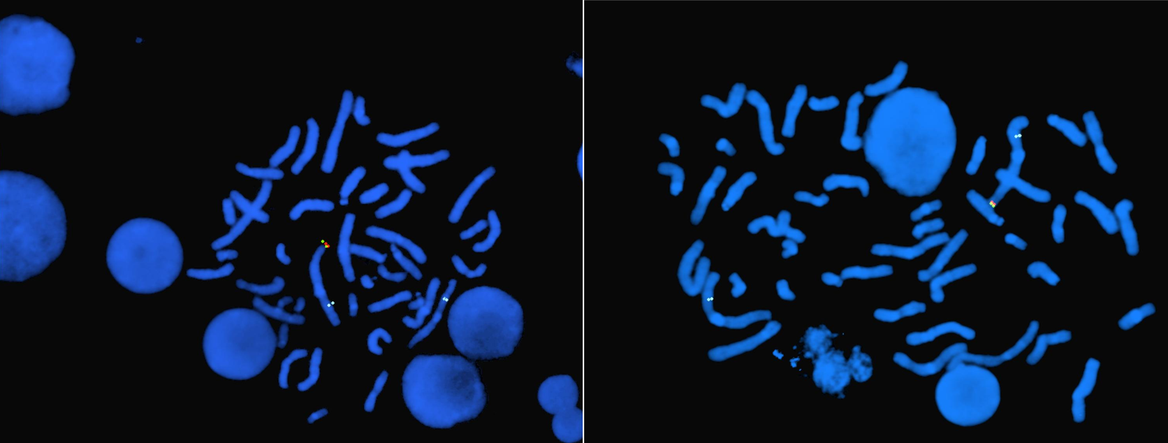
A total of 17 patients were included in the study. Four were diagnosed using microarray analysis, while the remaining patients were diagnosed using FISH following a preliminary diagnosis of 1p36 deletion syndrome (Figure 2). Among the cases diagnosed by microarray, three of four had no prior clinical suspicion, whereas one patient with a Robertsonian translocation, 45,XX,rob(13;14)(q10;q10), underwent microarray analysis despite having a preliminary diagnosis. Conventional karyotyping was performed in 14 patients, and a 1p36 deletion was detected in only one of them. The deletion size detected by microarray analysis ranged from 3685 kb to 15.7 Mb.
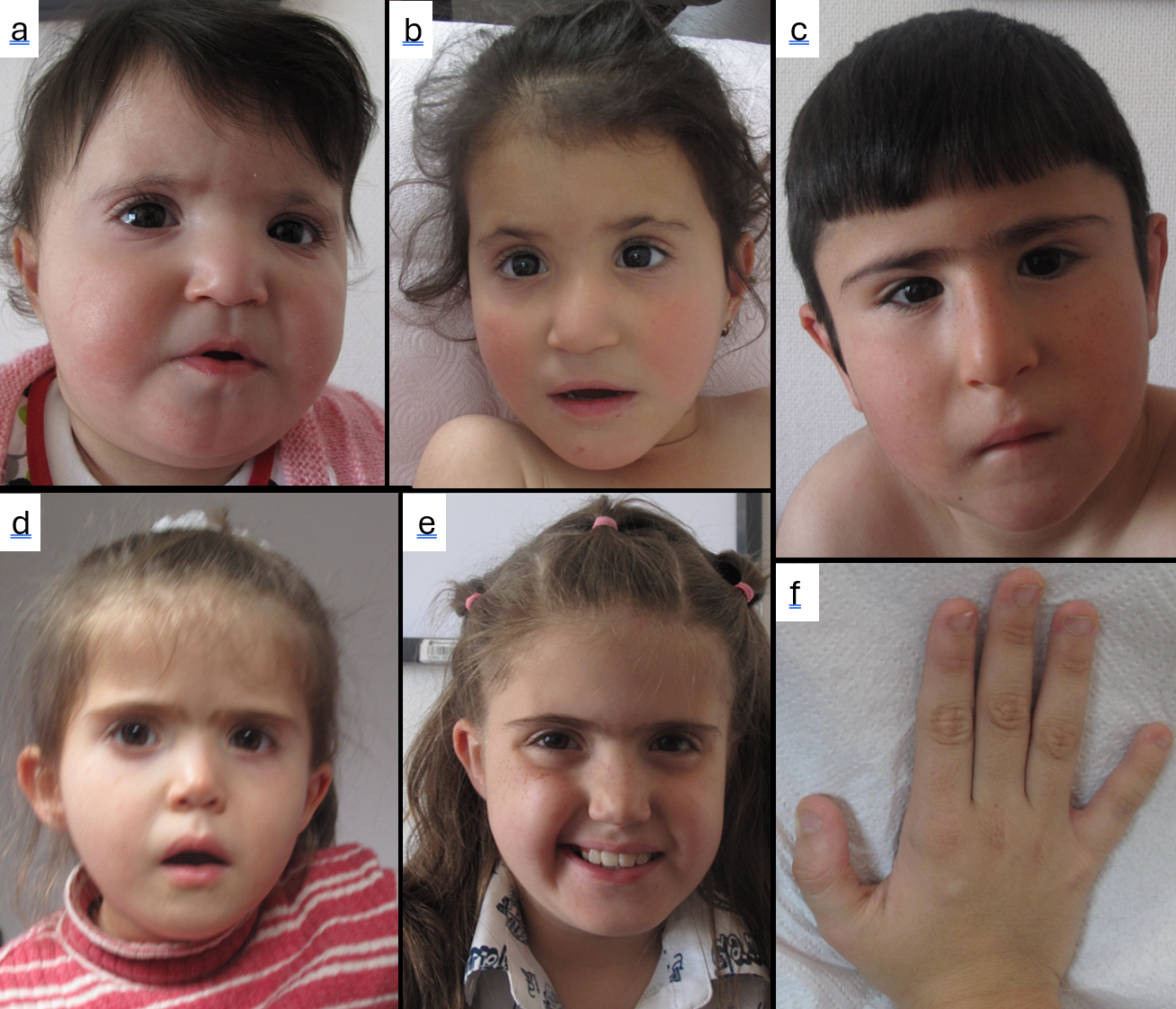
The mean age at admission and at diagnosis was 22 and 28 months, respectively. Prenatal CNS anomalies, including ventriculomegaly and corpus callosum hypoplasia, were detected in two patients. Growth impairment and microcephaly were common at presentation. Dysmorphic facial features were present in all patients, most commonly straight eyebrows, deep-set eyes, and ear abnormalities (Figure 1). Extremity abnormalities, including clubfoot, digital anomalies, and developmental hip dysplasia, were observed in eight patients.
Developmental delay was present in all patients. CNS abnormalities commonly included corpus callosum abnormalities, ventriculomegaly, and polymicrogyria. Congenital heart defects included atrial septal defect (ASD), ventricular septal defect (VSD), patent ductus arteriosus (PDA), bicuspid aortic valve, and aortic regurgitation, while noncompaction cardiomyopathy was also observed. Ophthalmological abnormalities included myopia, cataract, and optic disc pallor. Hearing loss, hypothyroidism, swallowing dysfunction, and cleft lip and palate were also identified.
Three of the 17 patients died, and three others could not be reached for long-term follow-up. All three children died of respiratory failure, two during infancy and one in the neonatal period. At the latest evaluation, severe neurodevelopmental impairment was evident among patients with available follow-up data. Only 4 of 11 patients (36.4%) achieved independent walking, and all did so at or after 24 months of age, while 9 of 11 patients (81.8%) were unable to perform daily activities without assistance. Also, language development was significantly affected, 4 of 11 patients (36.4%) remained non-verbal, while the others had a limited vocabulary (5–10 words) or were able to form only simple two-word sentences. Behavioral problems, particularly irritability, aggression, and autistic features, were detected in 5 of 11 patients (45.5%). Additionally, scoliosis was detected in 3 of 11 patients (27.3%) and obesity and/or hyperphagia in 4 of 11 patients (36.4%), with these findings becoming more pronounced after the age of 10. The demographic and clinical characteristics of the patients are summarized in Table 1.
| CNS: central nervous system, F: female, FISH: fluorescence in situ hybridization, M: male, SDS: standard deviation scores. | |
| Table 1. Summary of demographic characteristics, clinical findings, and diagnostic methods in patients with 1p36 deletion syndrome. | |
| Parameter |
|
| Demographic characteristics | |
| Sex (F/M) |
|
| Age at presentation (months) |
|
| Age at diagnosis (months) |
|
| Current age (years) |
|
| Maternal age at delivery (years) |
|
| Paternal age at delivery (years) |
|
| Diagnostic method | |
| FISH |
|
| Chromosomal microarray |
|
| Prenatal ultrasound abnormalities |
|
| Anthropometric measurements | |
| Height SDS |
|
| Weight SDS |
|
| Head circumference SDS |
|
| Short stature |
|
| Microcephaly |
|
| Facial dysmorphism | |
| Large anterior fontanel |
|
| Brachycephaly |
|
| Prominent forehead |
|
| Hypertelorism |
|
| Straight eyebrows |
|
| Deep-set eyes |
|
| Epicanthus |
|
| Depressed nasal bridge |
|
| Abnormal ears |
|
| Long philtrum |
|
| Micrognathia |
|
| Limb abnormalities |
|
| Developmental delay |
|
| CNS abnormalities |
|
| Epilepsy |
|
| Congenital heart defects |
|
| Cardiomyopathy |
|
| Ophthalmological abnormalities |
|
| Hearing loss |
|
| Hypothyroidism |
|
Discussion
Monosomy 1p36 is recognized as the most prevalent terminal chromosomal deletion in humans, which accounts for 0.5–1.2% of syndromic intellectual disability cases [7]. The phenotypic differences observed between affected individuals were initially thought to occur due to the parent-of-origin effect, but a study investigating this through DNA polymorphism analysis showed that there was no such effect [3]. The same study suggested that the size of 1p36 deletions varies significantly among patients and that these phenotypic differences may be due to deletions of varying sizes that result in the loss of different genes [3]. Moreover, differences in craniofacial features were not related solely to the size of the chromosomal deletion [3].
The deletion breakpoints and size show significant variability among patients and have been shown not to always correlate with the severity of the phenotype [9]. In a recent study, cases stratified into two groups as proximal or distal based on deletion location showed that cardiac abnormalities, CNS malformations, and epilepsy were more frequent in distal deletions, whereas microcephaly was more common in proximal deletions [5]. Additionally, deletions larger than 10 Mb have been shown to be associated with more severe neurodevelopmental outcomes [5]. In our study, due to the small number of patients in whom breakpoints were clearly identified by microarray analysis, it was not possible to establish such a correlation. However, microarray analysis in the two deceased patients showed deletions of different sizes, with a 15.7 Mb deletion in one and a 6922 kb deletion in the other. Conventional karyotyping detected the 1p36 deletion in only 1 of 14 patients tested in our cohort, indicating its limited diagnostic sensitivity for this syndrome. Therefore, when 1p36 deletion syndrome is clinically suspected, targeted FISH or CMA should be pursued even if the conventional karyotype is normal.
Previous studies suggest a female predominance; similarly, in our cohort, the female:male ratio was 13:4. More than 75% of patients exhibited characteristic dysmorphic facial features, including straight eyebrows, deep-set eyes, midface hypoplasia, a flat nasal bridge, posteriorly rotated, low‐set, and asymmetric ears, a long philtrum, and a pointed chin [7,10]. Additionally, microcephaly and a large or late-closing anterior fontanel are frequently observed. In our cohort, a characteristic facial gestalt was observed, particularly marked by straight eyebrows and deep-set eyes, similar to those described in the literature. In 14 out of 17 patients, we had a preliminary diagnosis, indicating that characteristic craniofacial features can effectively guide the diagnosis. Of the three patients for whom facial features did not initially lead to a diagnosis, one was evaluated as a newborn and the other two were around 2 years old. It was noted that dysmorphic features became more pronounced in patients over time. Haploinsufficiency of MMP23B (OMIM* 603321) has been proposed to contribute to a large or late-closing anterior fontanel, while SKI (OMIM* 164780) has been suggested as a candidate gene for the cleft lip/palate phenotype based on Ski-null mouse models [11,12].
Developmental delay and/or intellectual disability are observed in the vast majority of patients with 1p36 deletion syndrome, and haploinsufficiency of genes located within this locus, such as GNB1 (OMIM* 139380), CHD5 (OMIM* 610771), KCNAB2 (OMIM* 601142), SKI, GABRD (OMIM* 137163), MTOR (OMIM* 601231), RERE (OMIM* 605226), and CDC42 (OMIM* 116952), is thought to contribute to the neurodevelopmental phenotype [1,5]. All of our patients showed significant neurodevelopmental impairment based on clinical observations; however, formal cognitive assessment was not performed.
Epilepsy has been reported in 44–70% of cases, with both generalized and focal seizure types described, although infantile spasms are the most frequent [5,13,14]. Genotype–phenotype studies have shown that epilepsy is more frequent in distal deletions and that deletions involving GABRD and KCNAB2 are associated with the epilepsy phenotype [5]. Epilepsy was a prominent neurological feature in our cohort, observed in 70% of the patients.
CNS abnormalities, most notably ventriculomegaly and corpus callosum abnormalities, as well as cortical atrophy, periventricular nodular heterotopia, polymicrogyria, and white matter abnormalities, have been described [5,14-16]. In our cohort, consistent with the literature, ventriculomegaly and corpus callosum abnormalities were the most frequently observed findings. These anomalies may also be detectable during the prenatal period and should be kept in mind during evaluation.
Cardiac pathologies have been identified as the leading cause of mortality in the literature [5]. In the largest recent cohort, cardiovascular malformations or cardiomyopathy were reported in 47% of patients, including structural heart defects in 32% and cardiomyopathy in 21%, whereas the earlier Battaglia series reported congenital heart defects in 71% and cardiomyopathy in 27% [5,14]. While PRDM16 (OMIM*605557) and SKI have been proposed as the main genes implicated in cardiomyopathy, the relevant locus contains numerous genes, such as ECE1 (OMIM* 600423) and RERE, that are thought to contribute to the pathogenesis of cardiac defects [1,5]. HSPB7 (OMIM* 610692) has also been suggested as a potential candidate gene for cardiomyopathy based on population-level genetic association studies [17,18]. The frequency of congenital heart defects in our cohort was comparable to that reported in the largest recent cohort, whereas mortality was due to respiratory failure rather than cardiac causes. Among the patients who died, one had a large ASD, another had a bicuspid aortic valve with aortic regurgitation, and the third had no congenital heart defect; none showed cardiomyopathy.
Hypothyroidism has been reported, and screening for hypothyroidism is recommended at birth, at 6 months, and annually thereafter [10]. Hypothyroidism was observed in 4/8 (50%) of our patients, and since data for the remaining patients were unavailable, a precise frequency could not be determined; therefore, selection bias may exist.
Prader-Willi syndrome is included in the differential diagnosis, as some patients develop obesity after early feeding difficulties [19,20]. Among our patients, two were treated for swallowing dysfunction, and four developed obesity and/or hyperphagia during follow-up.
The main limitation of our study is that microarray analysis was not performed in all patients to precisely determine the breakpoints, which precluded the establishment of a genotype/phenotype correlation. Furthermore, due to the lack of parental studies, it could not be determined whether the deletion was de novo. Missing data for several clinical variables, particularly endocrine, ophthalmological, auditory, and CNS findings, led to varying denominators and may have affected the reported frequencies. Early diagnosis, along with timely screening and management of associated comorbidities, particularly neurological ones, is crucial for improving patient outcomes. Recognition of patients with a typical facial gestalt may facilitate earlier diagnosis and prevent unnecessary diagnostic testing. The possibility of balanced parental rearrangement should be considered, especially in cases with additional chromosomal copy number variations. Furthermore, germline mosaicism should be taken into account when providing genetic counseling to families regarding future pregnancies.
Ethical approval
This study was approved by the Hacettepe University Ethics Committee (Date: 23.03.2026, Decision/Protocol No: SBA 26/270). Informed consent was obtained from all participants involved in this study.
Data availability statement
The data that support the findings of this study are available from the corresponding author upon reasonable request.
Conflict of interest
The authors declare that this study was conducted in the absence of any commercial or financial relationships that could be construed as a potential conflict of interest.
Funding
The authors declare that this study received no funding.
Generative AI statement
The authors declare that no generative AI or AI-assisted technologies were used in the writing or preparation of this study.
References
- Jordan VK, Zaveri HP, Scott DA. 1p36 deletion syndrome: an update. Appl Clin Genet 2015;8:189-200. [Crossref]
- Hain D, Leversha M, Campbell N, Daniel A, Barr PA, Rogers JG. The ascertainment and implications of an unbalanced translocation in the neonate. Familial 1:15 translocation. Aust Paediatr J 1980;16(3):196-200. [Crossref]
- Shapira SK, McCaskill C, Northrup H, et al. Chromosome 1p36 deletions: the clinical phenotype and molecular characterization of a common newly delineated syndrome. Am J Hum Genet 1997;61(3):642-50. [Crossref]
- Gajecka M, Saitta SC, Gentles AJ, et al. Recurrent interstitial 1p36 deletions: Evidence for germline mosaicism and complex rearrangement breakpoints. Am J Med Genet A 2010;152A(12):3074-83. [Crossref]
- Jacquin C, Landais E, Poirsier C, et al. 1p36 deletion syndrome: review and mapping with further characterization of the phenotype, a new cohort of 86 patients. Am J Med Genet A 2023;191(2):445-58. [Crossref]
- Heilstedt HA, Ballif BC, Howard LA, Kashork CD, Shaffer LG. Population data suggest that deletions of 1p36 are a relatively common chromosome abnormality. Clin Genet 2003;64(4):310-6. [Crossref]
- Guterman S, Beneteau C, Redon S, et al. Prenatal findings in 1p36 deletion syndrome: new cases and a literature review. Prenat Diagn 2019;39(10):871-82. [Crossref]
- Akkuş PZ, Şahin Y, Utine E, Boduroğlu K. 1p36 microdeletion syndrome: a case report. Acta Medica. 2014;45(1):26-8.
- Rocha CF, Vasques RB, Santos SR, Paiva CLA. Mini-review: monosomy 1p36 syndrome: reviewing the correlation between deletion sizes and phenotypes. Genet Mol Res 2016;15(1):10.4238/gmr.15017942. [Crossref]
- Heilstedt HA, Ballif BC, Howard LA, et al. Physical map of 1p36, placement of breakpoints in monosomy 1p36, and clinical characterization of the syndrome. Am J Hum Genet 2003;72(5):1200-12. [Crossref]
- Colmenares C, Heilstedt HA, Shaffer LG, et al. Loss of the SKI proto-oncogene in individuals affected with 1p36 deletion syndrome is predicted by strain-dependent defects in Ski-/- mice. Nat Genet 2002;30(1):106-9. [Crossref]
- Gajecka M, Yu W, Ballif BC, et al. Delineation of mechanisms and regions of dosage imbalance in complex rearrangements of 1p36 leads to a putative gene for regulation of cranial suture closure. Eur J Hum Genet 2005;13(2):139-49. [Crossref]
- Bahi-Buisson N, Guttierrez-Delicado E, Soufflet C, et al. Spectrum of epilepsy in terminal 1p36 deletion syndrome. Epilepsia 2008;49(3):509-15. [Crossref]
- Battaglia A, Hoyme HE, Dallapiccola B, et al. Further delineation of deletion 1p36 syndrome in 60 patients: a recognizable phenotype and common cause of developmental delay and mental retardation. Pediatrics 2008;121(2):404-10. [Crossref]
- Descartes M, Mikhail FM, Franklin JC, McGrath TM, Bebin M. Monosomy1p36.3 and trisomy 19p13.3 in a child with periventricular nodular heterotopia. Pediatr Neurol 2011;45(4):274-8. [Crossref]
- Dobyns WB, Mirzaa G, Christian SL, et al. Consistent chromosome abnormalities identify novel polymicrogyria loci in 1p36.3, 2p16.1-p23.1, 4q21.21-q22.1, 6q26-q27, and 21q2. Am J Med Genet A 2008;146A(13):1637-54. [Crossref]
- Matkovich SJ, Van Booven DJ, Hindes A, et al. Cardiac signaling genes exhibit unexpected sequence diversity in sporadic cardiomyopathy, revealing HSPB7 polymorphisms associated with disease. J Clin Invest 2010;120(1):280-9. [Crossref]
- Stark K, Esslinger UB, Reinhard W, et al. Genetic association study identifies HSPB7 as a risk gene for idiopathic dilated cardiomyopathy. PLoS Genet 2010;6(10):e1001167. [Crossref]
- Tsuyusaki Y, Yoshihashi H, Furuya N, et al. 1p36 deletion syndrome associated with Prader-Willi-like phenotype. Pediatr Int 2010;52(4):547-50. [Crossref]
- D’Angelo CS, Da Paz JA, Kim CA, et al. Prader-Willi-like phenotype: investigation of 1p36 deletion in 41 patients with delayed psychomotor development, hypotonia, obesity and/or hyperphagia, learning disabilities and behavioral problems. Eur J Med Genet 2006;49(6):451-60. [Crossref]
Copyright and License
Copyright © 2026 The Author(s). This is an open access article distributed under the Creative Commons Attribution License (CC BY), which permits unrestricted use, distribution, and reproduction in any medium or format, provided the original work is properly cited.