


Abstract
Objective: Tibial plateau fractures are complex intra-articular injuries with variable functional outcomes. While fracture classifications guide management, their ability to predict recovery remains unclear. This study evaluated the impact of fracture severity, treatment variables, and fracture complexity on patient-reported and objective outcomes over time.
Methods: A retrospective cohort of 75 patients who underwent surgical treatment for tibial plateau fractures was analyzed. Functional outcomes were assessed using the Knee Injury and Osteoarthritis Outcome Score (KOOS) and knee flexion at 1 month, 3 months, 6 months, 1 year, and 2 years postoperatively. Fractures were classified using Schatzker and AO/OTA systems. Fixation method, column and segment involvement, and patient factors were recorded. Longitudinal changes were analyzed using generalized estimating equations, and predictors of early improvement were evaluated using robust regression.
Results: KOOS and knee flexion improved significantly over time (p<0.001). Mean KOOS increased from 23.15±5.87 at 1 month to 84.19±8.74 at 2 years, while flexion improved from 74.12±13.91° to 120.91±17.31°. Schatzker and AO/OTA classifications were associated with KOOS (p<0.001) but not flexion (p=0.160). Definitive internal fixation method was not significantly associated with KOOS outcomes (p=0.817). In robust regression, male sex (β=4.146, p=0.042) and posterior segment count (β=2.011, p=0.022) were positively associated with KOOS improvement, whereas comorbidity (β=−7.245, p=0.012) and provisional external fixation before definitive surgery (β=−8.683, p<0.001) were associated with less improvement, statistically.
Conclusion: Recovery following tibial plateau fractures is primarily time-dependent, with fracture severity influencing patient perception but not objective motion. No significant associations were identified between functional outcomes and fixation method or the evaluated fracture-complexity parameters in the present cohort.
Keywords: tibial plateau fractures, KOOS, fracture classification, range of motion, outcome assessment
Introduction
Tibial plateau fractures are complex intra-articular injuries that can significantly affect knee function and long-term joint health [1,2]. Restoration of articular congruity, mechanical alignment, and joint stability are considered essential for optimal recovery [1]. Despite advances in surgical techniques and fixation methods, functional outcomes remain variable, and the factors influencing recovery are not fully understood [2,3].
Fracture classification systems such as Schatzker and AO/OTA are widely used to describe injury patterns and guide treatment strategies [1,2]. In general, more severe fractures are thought to be associated with worse clinical outcomes [4,5]. At the same time, considerable emphasis has been placed on surgical technique, with the assumption that fixation method may influence postoperative recovery [3,6]. However, the extent to which fracture severity and fixation strategy independently affect functional outcomes remains unclear [1,2,4].
More recently, concepts such as the 3-column model and ten-segment classification have been introduced to better characterize fracture complexity and guide surgical planning [7-9]. Although these systems provide a more detailed anatomical description, their relationship with clinical outcomes has not been clearly established [8,9]. In addition, it is not well defined whether different outcome domains, such as patient-reported measures and objective range of motion, are influenced by the same factors [9,10].
Therefore, the aim of this study was to evaluate the impact of fracture severity, fixation method, and fracture complexity on functional outcomes following tibial plateau fractures. We hypothesized that fracture severity would negatively affect both patient-reported and objective outcomes, fixation method would have a significant impact on functional recovery.
Methods
The research was conducted in accordance with the principles of the Declaration of Helsinki. Ethical approval was obtained from the institutional review board (IRB No: SBA 25/682).
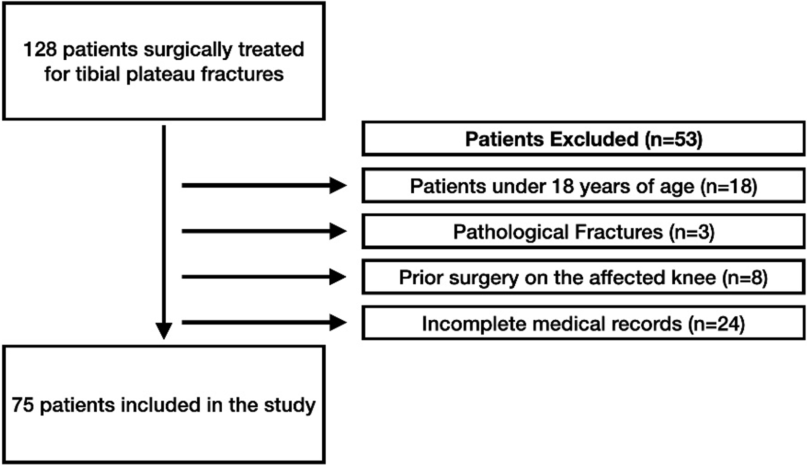
This retrospective cohort study included consecutive patients who underwent surgical treatment for tibial plateau fractures at a single tertiary referral center between 2014-2023. Patients were eligible for inclusion if they were aged ≥18 years, had a tibial plateau fracture treated surgically, and had available clinical and radiological follow-up data. Patients with pathological fractures, prior surgery on the affected knee, or incomplete medical records were excluded, Figure 1. All 75 included patients had available KOOS and knee flexion measurements at the predefined follow-up assessments.
Data collection
Demographic and clinical data were obtained from institutional records, including age at the time of surgery, sex, affected side, presence of comorbidities, trauma mechanism. Surgical variables included fixation method, use of bone graft (autograft, allograft, or none), application of external fixation, reoperation status. In the fixation classification, “plate plus screw(s)” indicated plate fixation combined with one or more additional independent lag screws placed outside the plate.
Fracture classification
Fractures were classified using preoperative imaging, including plain radiographs and computed tomography scans. Fracture patterns were categorized according to the Schatzker and AO/OTA classification systems [7-9]. Fracture complexity was further assessed using the 3-column concept and the ten-segment classification. The number of involved columns, as well as anterior and posterior segment involvement, was recorded for each patient.
Radiological assessment
Radiological measurements were performed using the institutional picture archiving and communication system (PACS). Preoperatively measured parameters included articular depression (mm), step-off (mm), plateau widening (mm), fracture gap (mm), varus/valgus alignment (degrees), and posterior tibial slope (degrees). All radiological measurements were performed by a single observer who was blinded to the clinical outcome data. Each parameter was measured independently on two separate occasions, and the mean of the two measurements was used for analysis.
Outcome measures
Functional outcomes were assessed using the validated Turkish version of the Knee Injury and Osteoarthritis Outcome Score (KOOS) and knee flexion range of motion measured in degrees. KOOS was calculated according to the standard scoring instructions, with higher scores indicating better knee-related outcomes. For the present analysis, an overall KOOS score was derived by calculating the global mean of the five individual validated subscale scores. Evaluations were performed at standardized postoperative time points: 1 month, 3 months, 6 months, 1 year, and 2 years [4,10-12]. Change scores for KOOS and knee flexion were calculated as the difference between the 2-year and 1-month postoperative values (2-year value minus 1-month value).
Statistical analysis
Statistical analyses were performed using IBM SPSS Statistics for Windows, Version 23.0 (IBM Corp., Armonk, NY, USA) and R software. Normality of continuous variables was assessed using the Kolmogorov–Smirnov test. Descriptive statistics were presented as mean ± standard deviation for normally distributed continuous variables, median (minimum–maximum) for non-normally distributed continuous variables, and frequency (percentage) for categorical variables. The effects of independent variables on non-normally distributed change scores were analyzed using robust regression analysis with the MASS package in R. To evaluate the effects of fixation method, Schatzker classification, AO/OTA classification, time, 3-column concept, posterior segment count, and anterior segment count on repeated KOOS and knee flexion measurements, generalized estimating equations (GEE) were used. Both main effects and time interactions were assessed in the GEE models. For variables with significant effects, multiple comparisons were performed using Bonferroni correction. For comparisons of parameters within the same time point based on the multi-response structure of the 3-column concept and ten-segment classifications, Bonferroni-adjusted multiple comparison tests were applied. Longitudinal changes over time were analyzed using generalized estimating equations (GEE), which account for within-subject correlations. A p-value <0.05 was considered statistically significant for all analyses.
Results
A total of 75 patients were included in the study, of whom 55 (73%) were male and 20 (27%) were female. The mean age at the time of surgery was 45.20±16.74 years. The most common trauma mechanism was fall (41%), followed by in-vehicle traffic accidents (33%). The right side was affected in 45% of patients. According to fracture classification, Schatzker type 6 (30%) and AO/OTA type 41-C (35%) fractures were the most frequent. Single plate with additional screw(s) was the most commonly used fixation method (31%). Bone graft was not used in 54.6% of cases. External fixation was applied in 19% of patients (Table 1). The complications were arthrofibrosis, infection, nonunion and implant failure.
| Data given as frequencies (percentages) or mean ± standard deviation. | ||
| Table 1. Demographic characteristics and clinical profile of patients. | ||
| Variables |
|
|
| Age |
|
|
| Sex | Male |
|
| Female |
|
|
| Side | Right |
|
| Left |
|
|
| Comorbidities | Present |
|
| None |
|
|
| Trauma Mechanism | Fall |
|
| In-Vehicle accident |
|
|
| Motorcycle accident |
|
|
| Pedestrian traffic accident |
|
|
| Scooter accident |
|
|
| Fixation Method | Single plate with additional screw(s) |
|
| Single Plate |
|
|
| Multiple plates with additional screw(s) |
|
|
| Screw fixation alone |
|
|
| Multiple Plates |
|
|
| Bone Graft Used | Autograft |
|
| Allograft |
|
|
| None |
|
|
| External Fixation Before Definitive Surgery |
|
|
| Reoperation Occurred |
|
|
| Schatzker Classification | 1 |
|
| 2 |
|
|
| 3 |
|
|
| 4 |
|
|
| 5 |
|
|
| 6 |
|
|
| AO/OTA Classification | 41-B1 |
|
| 41-B2 |
|
|
| 41-B3 |
|
|
| 41-C |
|
|
| Depression (mm) |
|
|
| Step-off (mm) |
|
|
| Plateau widening (mm) |
|
|
| Gap (mm) |
|
|
Both KOOS and knee flexion improved significantly over time. The mean KOOS increased from 23.15±5.87 at 1 month to 84.19±8.74 at 2 years (p<0.001). Similarly, mean knee flexion improved from 74.12±13.91° to 120.91±17.31° over the same period (p<0.001). Overall, improvements in both KOOS and knee flexion were statistically significant (p<0.001) (Table 2 and Table 3).
| Values are presented as mean ± standard deviation. Analyses were performed using generalized estimating equations. The reported p-values represent the overall effect of the corresponding variable in the GEE model. Different uppercase letters indicate statistically significant differences between groups at the same follow-up point, while different lowercase letters indicate statistically significant differences between follow-up points within the same group. Post hoc comparisons were adjusted using the Bonferroni method. A p-value <0.05 was considered statistically significant. | |||||||
| Table 2. KOOS outcomes. | |||||||
| Variables |
|
|
|
|
|
|
|
| Total KOOS Scores |
|
|
|
|
|
|
|
| Schatzker Classification | 1 |
|
|
|
|
|
|
| 2 |
|
|
|
|
|
||
| 3 |
|
|
|
|
|
||
| 4 |
|
|
|
|
|
||
| 5 |
|
|
|
|
|
||
| 6 |
|
|
|
|
|
||
| AO/OTA Classification | 41-B1 |
|
|
|
|
|
|
| 41-B2 |
|
|
|
|
|
||
| 41-B3 |
|
|
|
|
|
||
| 41-C |
|
|
|
|
|
||
| 3- Column | 1 column |
|
|
|
|
|
|
| 2 columns |
|
|
|
|
|
||
| 3 columns |
|
|
|
|
|
||
| Fixation Method | Single plate with additional screw(s) |
|
|
|
|
|
|
| Single plate |
|
|
|
|
|
||
| Multiple plates with additional screw(s) |
|
|
|
|
|
||
| Screw fixation alone |
|
|
|
|
|
||
| Multiple Plates |
|
|
|
|
|
||
| Affected Posterior Segment Count | 0 |
|
|
|
|
|
|
| 1 |
|
|
|
|
|
||
| 2 |
|
|
|
|
|
||
| 3 |
|
|
|
|
|
||
| 4 |
|
|
|
|
|
||
| 5 |
|
|
|
|
|
||
| Affected Anterior Segment Count | 0 |
|
|
|
|
|
|
| 1 |
|
|
|
|
|
||
| 2 |
|
|
|
|
|
||
| 3 |
|
|
|
|
|
||
| 4 |
|
|
|
|
|
||
| 5 |
|
|
|
|
|
||
| Values are presented as mean ± standard deviation. Analyses were performed using generalized estimating equations. The reported p-values represent the overall effect of the corresponding variable in the GEE model. Different uppercase letters indicate statistically significant differences between groups at the same follow-up point, while different lowercase letters indicate statistically significant differences between follow-up points within the same group. Post hoc comparisons were adjusted using the Bonferroni method. A p-value <0.05 was considered statistically significant. | |||||||
| Table 3. Knee flexion outcomes. | |||||||
| Variables |
|
|
|
|
|
|
|
| Total flexion range |
|
|
|
|
|
|
|
| Schatzker Classification | 1 |
|
|
|
|
|
|
| 2 |
|
|
|
|
|
||
| 3 |
|
|
|
|
|
||
| 4 |
|
|
|
|
|
||
| 5 |
|
|
|
|
|
||
| 6 |
|
|
|
|
|
||
| AO/OTA Classification | 41-B1 |
|
|
|
|
|
|
| 41-B2 |
|
|
|
|
|
||
| 41-B3 |
|
|
|
|
|
||
| 41-C |
|
|
|
|
|
||
| 3- Column | 1 column |
|
|
|
|
|
|
| 2 columns |
|
|
|
|
|
||
| 3 columns |
|
|
|
|
|
||
| Fixation Method | Single plate with additional screw(s) |
|
|
|
|
|
|
| Single plate |
|
|
|
|
|
||
| Multiple plates with additional screw(s) |
|
|
|
|
|
||
| Screw fixation alone |
|
|
|
|
|
||
| Multiple Plates |
|
|
|
|
|
||
| Affected Posterior Segment Count | 0 |
|
|
|
|
|
|
| 1 |
|
|
|
|
|
||
| 2 |
|
|
|
|
|
||
| 3 |
|
|
|
|
|
||
| 4 |
|
|
|
|
|
||
| 5 |
|
|
|
|
|
||
| Affected Anterior Segment Count | 0 |
|
|
|
|
|
|
| 1 |
|
|
|
|
|
||
| 2 |
|
|
|
|
|
||
| 3 |
|
|
|
|
|
||
| 4 |
|
|
|
|
|
||
| 5 |
|
|
|
|
|
||
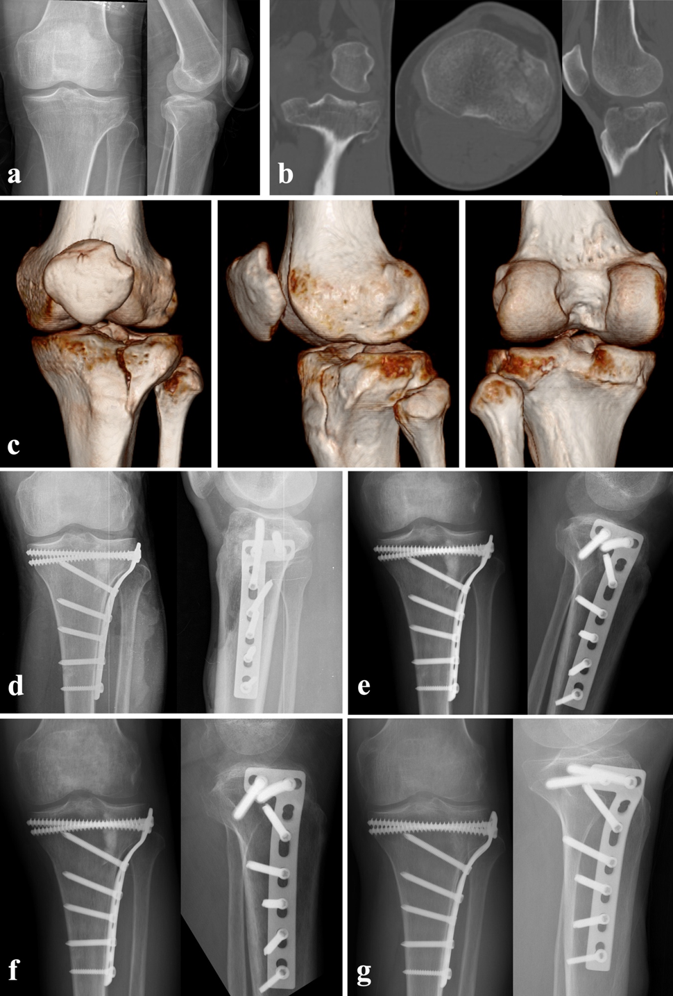
KOOS differed significantly according to fracture severity (p<0.001) (Table 2). In contrast, fixation method was not associated with differences in KOOS (p=0.817). Additionally, fracture complexity parameters, including the number of involved columns and anterior or posterior segment counts, were not associated with significant differences in overall KOOS values (p=0.243, p=0.316, and p=0.067, respectively) (Table 2). Representative preoperative, postoperative, and follow-up radiographs of a patient treated with plate-and-screw fixation are presented in Figure 2.
Knee flexion also improved significantly over time; however, no significant differences were observed between groups. Flexion values did not differ significantly according to Schatzker classification (p=0.160), AO/OTA classification (p=0.218), fixation method (p=0.882), or fracture complexity parameters (p>0.05 for all comparisons) (Table 3).
In the multivariable robust regression analysis, posterior segment count (β=2.011, 95% CI: 0.306–3.716, p=0.022) and male sex (β=4.146, 95% CI: 0.160–8.131, p=0.042) were positively associated with KOOS improvement. The presence of comorbidities (β=−7.245, 95% CI: −12.852 to −1.638, p=0.012) and the use of external fixation before definitive surgery (β=−8.683, 95% CI: −13.325 to −4.041, p<0.001) were associated with less improvement in KOOS (Table 4).
| β₁ represents the unstandardized regression coefficient with 95% confidence interval (CI), and β₂ represents the standardized regression coefficient. Analyses were performed using robust multivariable linear regression. Standard errors (SE), t-values, and p-values are reported for each predictor. Categorical variables were entered into the model using dummy coding, with the specified reference categories. A p-value < 0.05 was considered statistically significant. | ||||||
| Table 4. Multivariable robust regression analysis of independent predictors of change in KOOS score. | ||||||
| Variables |
|
|
|
|
|
|
| Constant |
|
|
|
|
|
|
| Age at surgery (year) |
|
|
|
|
|
|
| Male Sex |
|
|
|
|
|
|
| Presence of comorbidities |
|
|
|
|
|
|
| Articular surface depression (mm) |
|
|
|
|
|
|
| Articular step-off (mm) |
|
|
|
|
|
|
| Absolute plateau widening (mm) |
|
|
|
|
|
|
| Fracture gap (mm) |
|
|
|
|
|
|
| Fracture varus/valgus angle (°) |
|
|
|
|
|
|
| Posterior tibial slope (°) |
|
|
|
|
|
|
| Number of involved columns (3-column concept) |
|
|
|
|
|
|
| Number of involved posterior segments |
|
|
|
|
|
|
| Number of involved anterior segments |
|
|
|
|
|
|
|
Fixation method (reference: Single Plate) |
Single plate with additional screw(s) |
|
|
|
|
|
| Multiple Plates |
|
|
|
|
|
|
| Multiple plates with additional screw(s) |
|
|
|
|
|
|
| Screw fixation alone |
|
|
|
|
|
|
| Graft use (reference: none) | Allograft |
|
|
|
|
|
| Autograft |
|
|
|
|
|
|
| External fixation occurred before definitive surgery |
|
|
|
|
|
|
| Reoperation occurred |
|
|
|
|
|
|
Discussion
In this study, both patient-reported outcomes and objective knee motion demonstrated significant improvement over time following surgical treatment of tibial plateau fractures. However, important differences emerged when outcomes were analyzed in relation to fracture characteristics. Both the AO/OTA and Schatzker classifications were significantly associated with KOOS scores (p<0.001 for both), whereas neither classification showed a statistically significant association with knee flexion (p=0.218 and p=0.160, respectively). In contrast, fixation method and fracture complexity, including column and segment involvement, were not associated with either functional or objective outcomes. These findings suggest that recovery after tibial plateau fractures is multifactorial and not solely determined by fracture morphology or surgical technique.
The progressive improvement observed in both KOOS and knee flexion over time is consistent with previous literature. Lang et al. 2025 demonstrated that longer follow-up duration is associated with significantly better patient-reported outcomes, reflecting ongoing biological healing and functional adaptation [10]. Similarly, several longitudinal cohort studies have shown that functional recovery continues beyond the early postoperative period, often extending up to two years and even up to five years in some domains [13,14]. However, certain basic outcomes such as range of motion may plateau earlier, typically between 6 and 12 months [15]. Our findings support this pattern, emphasizing that recovery following tibial plateau fractures follows a gradual and domain-dependent trajectory rather than reaching an early universal plateau.
Fracture severity remains an important determinant of outcome; however, its effect appears to be domain specific. In the present study, KOOS scores differed significantly according to both the Schatzker and AO/OTA classifications, whereas knee flexion did not differ significantly between classification groups. This is consistent with previous reports showing that complex fracture patterns (Schatzker IV–VI) are associated with worse patient-reported outcomes, particularly in KOOS sports and QoL domains [10]. However, objective range of motion may remain comparable across fracture severities, as demonstrated in mid-term follow-up cohorts reporting no significant differences in knee flexion between simple and complex fractures [16]. In addition, Madi et al. demonstrated that greater postoperative articular step-off is associated with worse patient-reported outcomes, while Sadighi et al. reported that increased depression correlates with inferior clinical scores, with minimal impact on ROM [5,17]. Our findings extend this knowledge by demonstrating that the impact of fracture severity may be more pronounced in patient perception than in objective range of motion, suggesting that different outcome domains capture distinct aspects of recovery.
The discrepancy between patient-reported outcomes and objective knee motion represents one of the most important findings of this study. While KOOS scores were influenced by fracture severity, knee flexion was not, indicating that subjective and objective measures of recovery do not necessarily align. This observation is supported by previous literature. Elsoe et al. found that MRI-verified soft tissue injuries did not significantly affect KOOS scores at one year, highlighting the complex and multifactorial nature of patient-reported outcomes [18]. Likewise, Vaartjes et al. demonstrated in a large multicenter cohort that although many patients regain functional independence in pain and ADL domains, deficits in KOOS sports and QoL often persist, particularly in higher-demand activities [3]. These findings suggest that pain, perceived instability, activity modification, and psychosocial factors play a critical role in subjective recovery. Furthermore, mental health status has been shown to significantly influence long-term outcomes after tibial plateau fractures [19]. Therefore, evaluation of outcomes after tibial plateau fractures should incorporate both subjective and objective measures to provide a comprehensive assessment.
Another notable finding of this study is that no statistically significant association between fixation method and clinical outcomes was identified in the present cohort. This aligns with current evidence suggesting that surgical technique may be less important than achieving adequate reduction and stability. A recent meta-analysis by Tay et al. found no significant differences in functional outcomes or complication rates between arthroscopy-assisted and conventional open reduction techniques [20]. While some studies, such as van der Linden et al., have suggested that greater fracture extent may be associated with worse patient-reported outcomes, the overall prognostic value of classification systems remains limited [21]. This is further supported by systematic reviews highlighting the variability and moderate reliability of commonly used classification systems, as well as their limited ability to predict clinical outcomes [22]. Taken together, these findings suggest that although classification systems remain valuable for describing fracture morphology and guiding surgical planning, their additional prognostic value remains debatable.
The regression analysis in this study further supports the multifactorial nature of recovery. Comorbidities were associated with worse early functional improvement, which is consistent with existing literature demonstrating the negative impact of patient-related factors on recovery. Studies evaluating clinical outcomes and quality of life after tibial plateau fractures have shown that factors such as increased body weight and general health status negatively influence both physical and psychological recovery [23]. Additionally, injury-related factors such as polytrauma and fracture complexity have been associated with reduced range of motion at one year [4]. Interestingly, posterior segment involvement was associated with greater early improvement in our cohort; however, this finding contrasts with some previous reports suggesting worse high-demand functional outcomes and limited sports participation in such cases, often due to pain and fear of reinjury [24]. Therefore, this result should be interpreted cautiously. The use of external fixation before definitive surgery was also associated with less early improvement in KOOS. However, external fixation is generally used in patients with more severe injuries or greater soft-tissue compromise. Therefore, this association may reflect the initial injury severity rather than an independent adverse effect of external fixation itself. Overall, these findings underscore the importance of considering both patient-related and injury-related factors when evaluating prognosis and reinforce the need for a multidimensional assessment of recovery.
This study has several limitations. First, its retrospective design introduces the potential for selection bias. Second, all radiographic measurements were performed by a single observer, and interobserver reliability was not assessed. Third, stratifying our 75 patients into multiple classification and fixation categories created small subgroups. This may have introduced statistical power constraints and a risk of Type II errors. Fourth, the cohort was heterogeneous with respect to fracture pattern, associated injuries and treatment, and the fixation method was selected by the treating surgeon according to fracture morphology and soft-tissue condition rather than at random; residual confounding by indication therefore cannot be excluded. Fifth, because several subgroups were small, some statistically significant associations may not be clinically meaningful and should be regarded as exploratory; larger, preferably prospective studies are needed to confirm these findings.
Conclusion
Functional recovery after surgically treated tibial plateau fractures continued throughout the two-year follow-up period. Fracture severity was associated with patient-reported KOOS outcomes but not with knee flexion, statistically. No statistically significant associations between the evaluated fixation methods or fracture-complexity parameters and functional outcomes were identified in this cohort. These findings support the combined use of patient-reported and objective measures when evaluating recovery after tibial plateau fractures.
Ethical approval
This study was approved by the Hacettepe Üni̇versi̇tesi̇ Sağlik Bi̇li̇mleri̇ Araştirma Eti̇k (Date: 22 July 2025, IRB No: SBA 25/682). Informed consent was obtained from all participants involved in this study.
Data availability statement
The data that support the findings of this study are available from the corresponding author upon reasonable request.
Conflict of interest
The authors declare that this study was conducted in the absence of any commercial or financial relationships that could be construed as a potential conflict of interest.
Funding
The authors declare that this study received no funding.
Generative AI statement
The authors declare that no generative AI or AI-assisted technologies were used in the writing or preparation of this study.
References
- Prat-Fabregat S, Camacho-Carrasco P. Treatment strategy for tibial plateau fractures: an update. EFORT Open Rev 2017;1(5):225-32. https://doi.org/10.1302/2058-5241.1.000031
- Schatzker J, Kfuri M. Revisiting the management of tibial plateau fractures. Injury 2022;53(6):2207-18. https://doi.org/10.1016/j.injury.2022.04.006
- Vaartjes TP, van der Sluis FJ, Bosma E, et al. Most patients with tibial plateau fractures regain function for daily activities but do not return to their pre-injury level of sport: comparison of multicenter cohort of 1101 patients and age-related peers. Eur J Trauma Emerg Surg 2025;51(1):285. https://doi.org/10.1007/s00068-025-02958-9
- Li J, Zhang J, Zhao K, et al. Incidence and risk factors for decreased range of motion of the knee joint after surgery for closed tibial plateau fracture in adults. J Orthop Surg Res 2021;16(1):549. https://doi.org/10.1186/s13018-021-02700-2
- Madi S, Chotta S, Fadel PA, Fares MY, Zeichen J. Association between articular surface depression and functional outcomes after ORIF of tibial plateau fractures: a retrospective cohort study. Eur J Trauma Emerg Surg 2025;51(1):357. https://doi.org/10.1007/s00068-025-03027-x
- Urruela AM, Davidovitch R, Karia R, Khurana S, Egol KA. Results following operative treatment of tibial plateau fractures. J Knee Surg 2013;26(3):161-5. https://doi.org/10.1055/s-0032-1324807
- Gahlot N, Kunal K, Elhence A, et al. Ten-segment classification has lowest inter/intra-observer reliability as compared to schatzker, three-column and ao systems for tibial plateau fractures: a comparison based on surgeons’ experience. Arch Bone Jt Surg 2023;11(4):256-61. https://doi.org/10.22038/ABJS.2022.63442.3061
- Koksal A, Abul K, Kargın D, Cimen O, Mert M, Keskin A. An evaluation of intra and inter observer reliability of the five used classification systems of tibial plateau fractures. Acta Orthop Belg 2022;88(4):805-10. https://doi.org/10.52628/88.4.9845
- Vosoughi F, Menbari Oskouie I, Rahimdoost N, Pesantez R. Decoding tibial plateau fracture classifications: a century of individualized insights in a systematic review. EFORT Open Rev 2025;10(5):316-26. https://doi.org/10.1530/EOR-2024-0184
- Lang C, Hörmandinger C, Klaut M, et al. Clinical outcome in tibial plateau fractures improves over time: Insights from a collaborative data network. Injury 2025;56(8):112607. https://doi.org/10.1016/j.injury.2025.112607
- Paker N, Buğdaycı D, Sabırlı F, Özel S, Ersoy S. Knee injury and osteoarthritis outcome score: reliability and validation of the Turkish version. Turkiye Klinikleri J Med Sci. 2007;27(3):350-6.
- Roos EM, Roos HP, Lohmander LS, Ekdahl C, Beynnon BD. Knee Injury and Osteoarthritis Outcome Score (KOOS)-development of a self-administered outcome measure. J Orthop Sports Phys Ther 1998;28(2):88-96. https://doi.org/10.2519/jospt.1998.28.2.88
- Gonzalez LJ, Hildebrandt K, Carlock K, Konda SR, Egol KA. Patient function continues to improve over the first five years following tibial plateau fracture managed by open reduction and internal fixation. Bone Joint J 2020;102-B(5):632-7. https://doi.org/10.1302/0301-620X.102B5.BJJ-2019-1385.R1
- Millar SC, Bennett K, Fraysse F, Arnold JB, Solomon LB, Thewlis D. Longitudinal changes in lower limb joint loading up to two years following tibial plateau fracture. Gait Posture 2020;78:72-9. https://doi.org/10.1016/j.gaitpost.2020.03.008
- Christiano AV, Pean CA, Kugelman DN, Konda SR, Egol KA. Function and knee range of motion plateau six months following lateral tibial plateau fractures. J Knee Surg 2020;33(5):481-5. https://doi.org/10.1055/s-0039-1678676
- Neidlein C, Watrinet J, Pätzold R, et al. Patient-reported outcomes following tibial plateau fractures: mid- to short-term implications for knee function and activity level. J Clin Med 2024;13(8):2327. https://doi.org/10.3390/jcm13082327
- Sadighi M, Sabaghzadeh A, Biglari F, Ebrahimpour A, Karami A, Jafari Kafiabadi M. Clinical outcomes of Schatzker type II tibial plateau fractures using joint depression morphology: a cross-sectional study. Acta Orthop Belg 2023;89(1):128-34. https://doi.org/10.52628/89.1.10846
- Elsoe R, Motahar I, Mahdi F, Larsen P. Presence of magnetic resonance imaging verified soft tissue injuries did not significantly affect the patient-reported outcome 12 months following a lateral tibial plateau fracture: a 12-month prospective cohort study of 56 patients. Knee 2020;27(2):420-7. https://doi.org/10.1016/j.knee.2020.01.008
- Kugelman D, Qatu A, Haglin J, Konda S, Egol K. Impact of psychiatric illness on outcomes after operatively managed tibial plateau fractures (OTA-41). J Orthop Trauma 2018;32(6):e221-5. https://doi.org/10.1097/BOT.0000000000001138
- Tay ST, Chen MZ, Chan YS, Kuo LT. The efficacy of arthroscopy-assisted versus stand-alone open reduction and internal fixation for treating tibial plateau fracture: a systematic review and meta-analysis. BMC Musculoskelet Disord 2024;25(1):865. https://doi.org/10.1186/s12891-024-07958-1
- van der Linden LR, Vaartjes TP, Kramer CJSA, et al. What is the impact of the fracture location on patient-reported functional outcomes in patients with lateral tibial plateau fractures? Injury 2025;56(11):112720. https://doi.org/10.1016/j.injury.2025.112720
- Millar SC, Arnold JB, Thewlis D, Fraysse F, Solomon LB. A systematic literature review of tibial plateau fractures: what classifications are used and how reliable and useful are they? Injury 2018;49(3):473-90. https://doi.org/10.1016/j.injury.2018.01.025
- Höller LF, Höller S, Klinger E, et al. Clinical outcomes and quality of life of patients after surgical treatment of a tibia plateau fracture. Eur J Trauma Emerg Surg 2025;51(1):319. https://doi.org/10.1007/s00068-025-02963-y
- Quintens L, Van den Berg J, Reul M, et al. Poor sporting abilities after tibial plateau fractures involving the posterior column: how can we do better? Eur J Trauma Emerg Surg 2021;47(1):201-9. https://doi.org/10.1007/s00068-019-01220-3
Copyright and License
Copyright © 2026 The Author(s). This is an open access article distributed under the Creative Commons Attribution License (CC BY), which permits unrestricted use, distribution, and reproduction in any medium or format, provided the original work is properly cited.