


Abstract
Objective: Alterations in mismatch repair (MMR) protein expression have been reported in liver cancers, including hepatocellular carcinoma and cholangiocarcinoma. Loss or reduced expression of MMR proteins is commonly associated with microsatellite instability (MSI), which has important prognostic and therapeutic implications. Immunohistochemistry represents a cost-effective and reliable method for detecting deficient MMR (dMMR). However, due to potential technical pitfalls and variable staining patterns, careful interpretation of MMR immunostaining is required.
Materials and Methods: In this study, virtual slides of tumor microarray cores from 35 patients were re-evaluated. The cases were stained with different antibody clones targeting MLH1, PMS2, MSH2, and MSH6. Based on the percentage of nuclear staining, immunohistochemical expression patterns were categorized as: (1) total expression loss (<25% nuclear staining), (2) focal expression loss (25-75% nuclear staining), and (3) intact expression (>75% nuclear staining).
Results: Among 35 cases (21 hepatocellular carcinomas and 14 cholangiocarcinomas), clone-dependent variability in MMR immunohistochemistry performance was observed. Using the most effective antibody clones, potential dMMR patterns in hepatocellular carcinoma were identified in 25% of cases for MLH1, 50% for MSH2, and 50% for MSH6. In contrast, no dMMR-like pattern was observed in cholangiocarcinoma for any MMR protein. PMS2 (single available clone, CAB010235) demonstrated minimal expression in normal liver tissue and variable staining in tumors, suggesting technical limitations rather than true biological loss. These findings indicate that suboptimal antibody clones may artificially increase the apparent dMMR rate.
Conclusion: This study evaluated MMR protein expression patterns in hepatocellular carcinoma and cholangiocarcinoma using data from the Human Protein Atlas, an open-access resource. Although immunohistochemistry remains a cost-effective and reliable method, factors such as fixation quality, antibody clone sensitivity, and careful evaluation of internal controls must be considered. Appropriate antibody clone selection is therefore essential for accurate interpretation of MMR immunostaining.
Keywords: MMR, liver, Immunohistochemistry, Cancer, Human Protein Atlas
Introduction
Mismatch repair (MMR) proteins play a critical role in maintaining genomic stability by correcting DNA replication errors [1,2]. Loss of function of at least one of the four key MMR proteins—MLH1, PMS2, MSH2, or MSH6—leads to impairment of the DNA mismatch repair system and results in microsatellite instability (MSI-H), a hypermutable condition observed in various tumor types. Loss of MMR protein expression is therefore commonly associated with MSI-H, a genomic instability phenotype that may arise from either somatic or germline alterations [3].
The frequency of deficient MMR (dMMR) or MSI-H in liver tumors varies according to tumor type and histologic grade [4-7]. In well-differentiated hepatocellular carcinomas (grade 1-2), the frequency of dMMR/MSI-H is generally lower than that observed in poorly differentiated tumors (grade 3), likely reflecting lower proliferative activity and more preserved DNA repair capacity [8].
Several methods, including immunohistochemistry (IHC), polymerase chain reaction (PCR), and next-generation sequencing (NGS), are used to evaluate MMR deficiency in tumors. Among these, immunohistochemistry is a widely used, cost-effective, and practical method for detecting MMR dysfunction [9-11]. However, this technique has several technical limitations and interpretative challenges that require expertise in evaluating staining patterns in both normal and neoplastic tissues. Importantly, MMR protein staining patterns may vary among different tissues, tumor types, and even within the same tumor. In addition, expression of MMR proteins is highly sensitive to tissue fixation conditions [12]. Focal loss of staining may arise from tumor subclones with MMR dysfunction, but it may also result from fixation-related artifacts or abnormal genetic alterations, such as novel mutations or aberrant cytoplasmic accumulation of mutant proteins [13,14].
The present study aims to highlight the technical and interpretative challenges associated with MMR immunohistochemistry, with particular emphasis on antibody clone characteristics, tissue fixation quality, and the role of pathologist expertise. Liver tumors were selected as a model because hepatocellular carcinoma and cholangiocarcinoma display distinct biological and proliferative characteristics. These differences provide an opportunity to demonstrate clone- and tissue-dependent variability in MMR staining patterns and the potential risk of misinterpretation, particularly in well-differentiated, low-proliferation tumors such as hepatocellular carcinoma. Using data derived from the Human Protein Atlas, we evaluated MMR staining patterns across different antibody clones and tissue types in order to emphasize the importance of internal controls and technical awareness for accurate interpretation of MMR immunohistochemistry.
Materials and methods
Study cohort and tissue microarray (tma) core selection
Adult patients with hepatocellular carcinoma or cholangiocarcinoma whose immunohistochemical tissue microarray (TMA) data were available in the open-access Human Protein Atlas (HPA) database were included in the study cohort [15-17].
Immunohistochemical staining for proteins encoded by four MMR genes—MLH1 (clone 1: CAB013294, clone 2: HPA052707, clone 3: CAB070868), PMS2 (clone 1: CAB010235), MSH2 (clone 1: CAB009572, clone 2: CAB070867, clone 3: CAB080131), and MSH6 (clone 1: CAB009091, clone 2: HPA028376, clone 3: HPA028446, clone 4: CAB070870)—was evaluated in the available TMA cores. Normal liver tissue cores were also assessed as controls.
Clinical information including tumor type, age, and sex was obtained from the Human Protein Atlas database (Supplementary Table 1 and Supplementary Table 2) (https://www.proteinatlas.org, accessed August 25 2023).
Evaluation of protein-level expression
Evaluation of MMR protein expression in normal liver tissue cores: In normal liver tissue cores, nuclear staining in hepatocytes, cholangiocytes, fibroblasts, and inflammatory cells was considered the expected staining pattern (0 = loss, 1 = intact) (Figure 1). These cell populations were also used as internal controls for the evaluation of tumor tissues (Supplementary Table 1 and Supplementary Table 2).
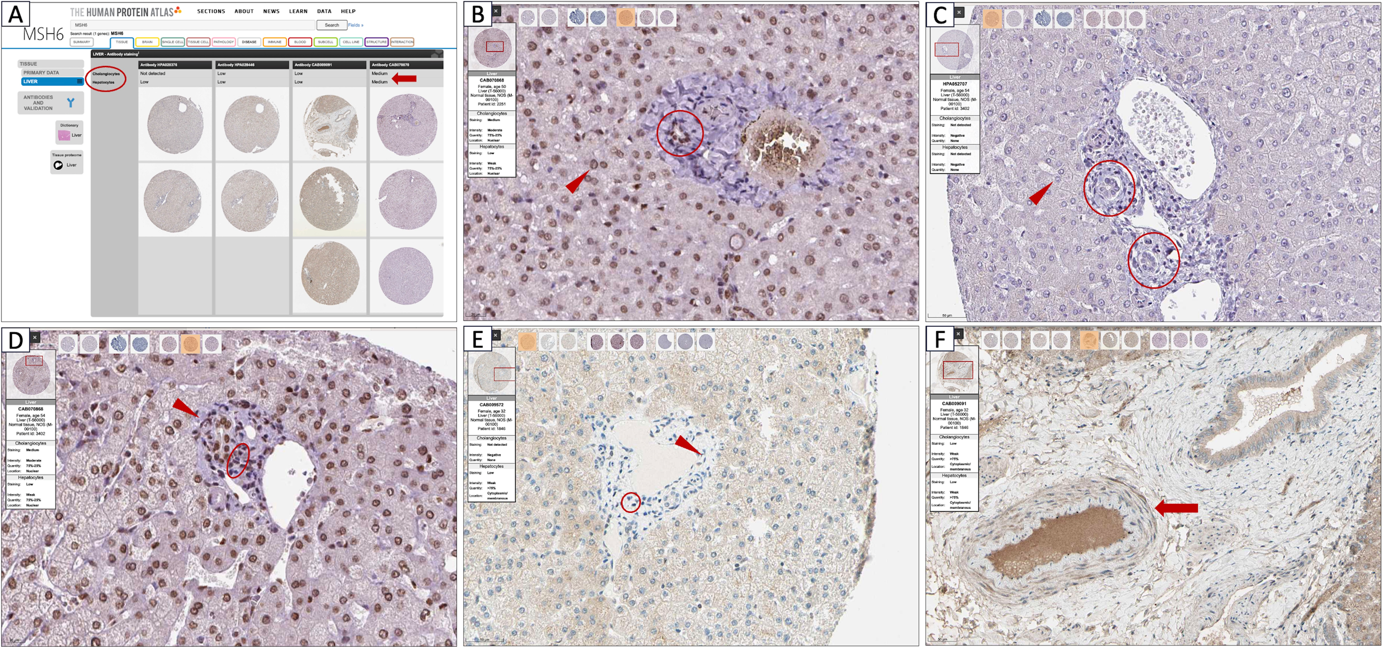
Evaluation of MMR protein expression in neoplastic cores: In tumor cores, peritumoral stromal cells—including hepatocytes, cholangiocytes, fibroblasts, and inflammatory cells—were assessed as internal controls based on their nuclear staining status (0 = loss, 1 = intact).
To distinguish fixation-related staining defects from clone-related staining variability, each core was evaluated using a modified H-score calculated by multiplying the percentage of positive cells by staining intensity (0-3 scale: 0 = negative, 1 = weak, 2 = moderate, 3 = strong) (Supplementary Table 1).
Based on the percentage of tumor cells showing nuclear staining, immunohistochemical expression patterns were categorized as: 1) Total expression loss (<25% nuclear staining), 2) Focal expression loss (25-75% nuclear staining) and 3) Intact expression (>75% nuclear staining). These categories corresponded to the following MMR status groups:
1.dMMR (deficient MMR): Loss of nuclear protein expression of at least one MMR protein (MLH1, PMS2, MSH2, or MSH6) in tumor cells while internal control cells retained intact staining (Figure 2).
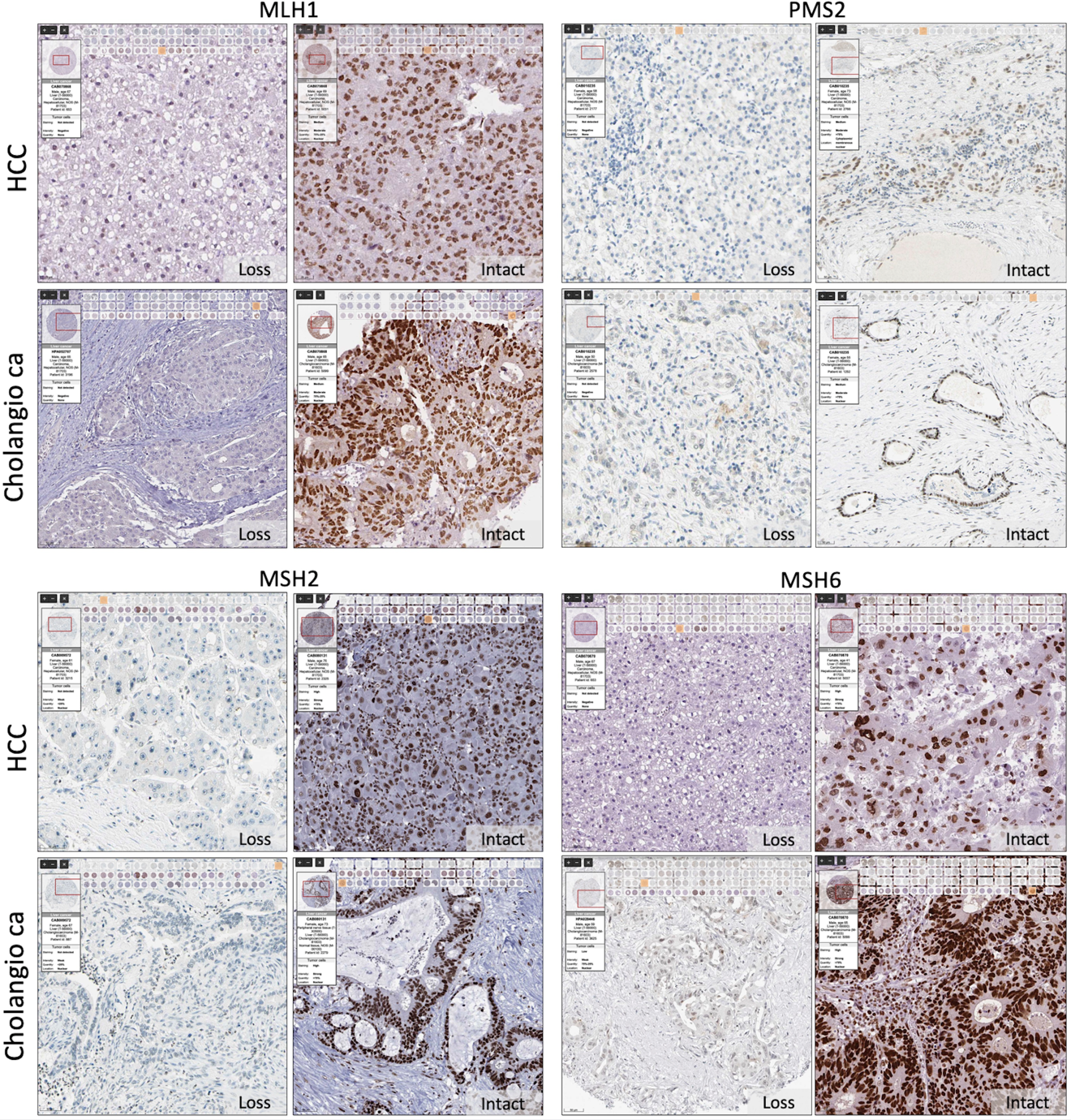
MLH1 – Loss: Patient ID 2767 (M, 58 y, liver hepatocellular carcinoma, clone CAB013294); Intact: Patient ID 3402 (F, 54 y, liver hepatocellular carcinoma, clone CAB013294). PMS2 – Loss: Patient ID 2251 (F, 50 y, liver cholangiocarcinoma, clone CAB010235); Intact: Patient ID 3402 (F, 54 y, liver cholangiocarcinoma, clone CAB010235). MSH2 – Loss: Patient ID 1846 (F, 32 y, liver hepatocellular carcinoma, clone CAB080131); Intact: Patient ID 2251 (F, 50 y, liver cholangiocarcinoma, clone CAB080131). MSH6 – Loss: Patient ID 1846 (F, 32 y, liver hepatocellular carcinoma, clone CAB070870); Intact: Patient ID 2251 (F, 50 y, liver cholangiocarcinoma, clone CAB070870).
HCC = hepatocellular carcinoma; Cholangio ca = cholangiocarcinoma; Loss = absence of nuclear staining in tumor cells; Intact = retained nuclear staining in tumor cells. Scale bars, dataset identifiers, and clone codes are displayed on each panel.
2a.fdMMR (focal deficient MMR): Loss of nuclear staining in a subset of tumor cells, reflecting intratumoral heterogeneity, with preserved staining in surrounding internal control cells.
2b.Fixation-related staining loss: Concurrent loss of staining in both tumor cells and surrounding internal control cells, suggesting technical artifact rather than true biological loss.
3.pMMR (proficient MMR): Preserved nuclear staining of all four MMR proteins (MLH1, PMS2, MSH2, and MSH6) in tumor cells (Figure 2).
Results
Demographic findings - All individuals included in the study were adults, including those contributing normal liver tissue used as controls. The mean age of the patients (n=35) was 63.62 years (range: 24-82 years). Among the patients, 21 had hepatocellular carcinoma and 14 had cholangiocarcinoma. A summary of the demographic characteristics is provided in Table 1.
| HCC: Hepatocellular carcinoma, Cholangio ca: Cholangiocarcinoma. | |||
| Table 1. Patients’ demographic data | |||
|
|
|
|
|
| Number of patients (n) |
|
|
|
| Age (year) | |||
| Mean |
|
|
|
| Min |
|
|
|
| Max |
|
|
|
| Gender | |||
| Female |
|
|
|
| Male |
|
|
|
Clone dependent antibody evaluation results
MLH1 - Based on the evaluation of staining patterns in control tissues, clone 3 was identified as the most effective antibody clone (Figure 3, Supplementary Table 3). In all normal liver tissue cores, hepatocytes, cholangiocytes, fibroblasts, and inflammatory cells showed nuclear positivity for MLH1, indicating intact expression. In neoplastic tissues, both hepatocellular carcinomas and cholangiocarcinomas also demonstrated preserved staining in internal control cells (Figure 4 and Figure 5). Among hepatocellular carcinomas, one tumor showed total expression loss (<25% nuclear staining) and one tumor demonstrated focal expression loss (25-75% nuclear staining), whereas six of the eight tumors showed intact expression (>75% nuclear staining). In cholangiocarcinomas, all four tumors showed intact expression. No cases of total or focal expression loss were observed.
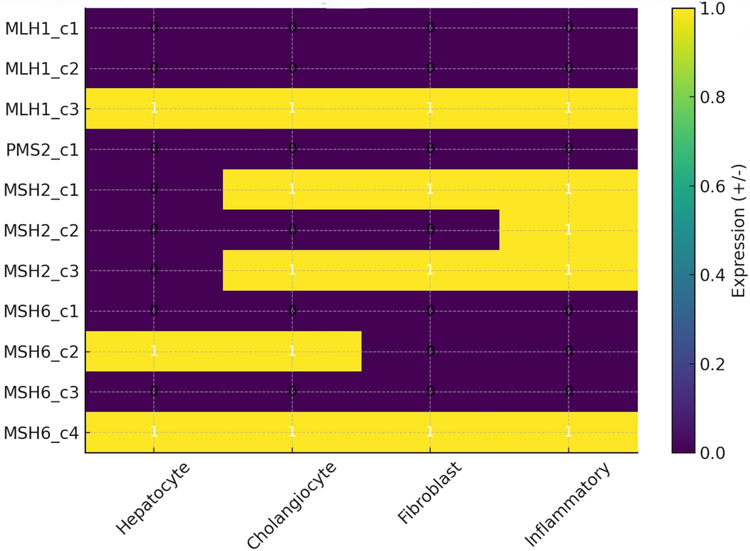
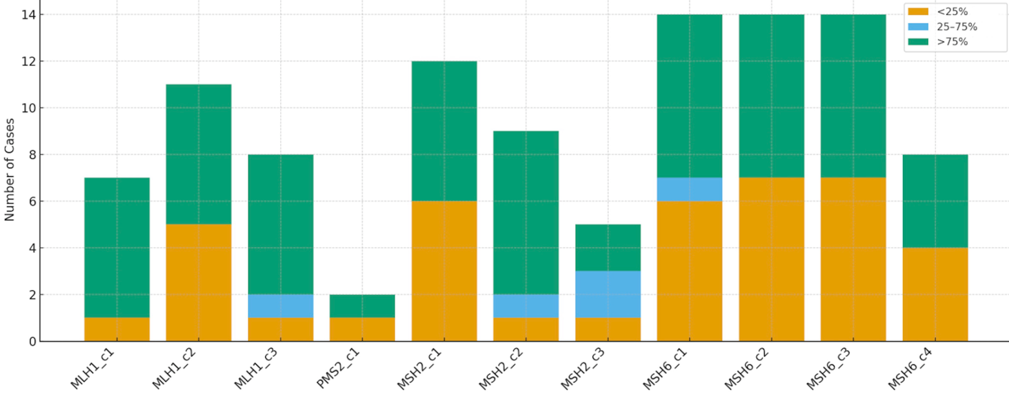
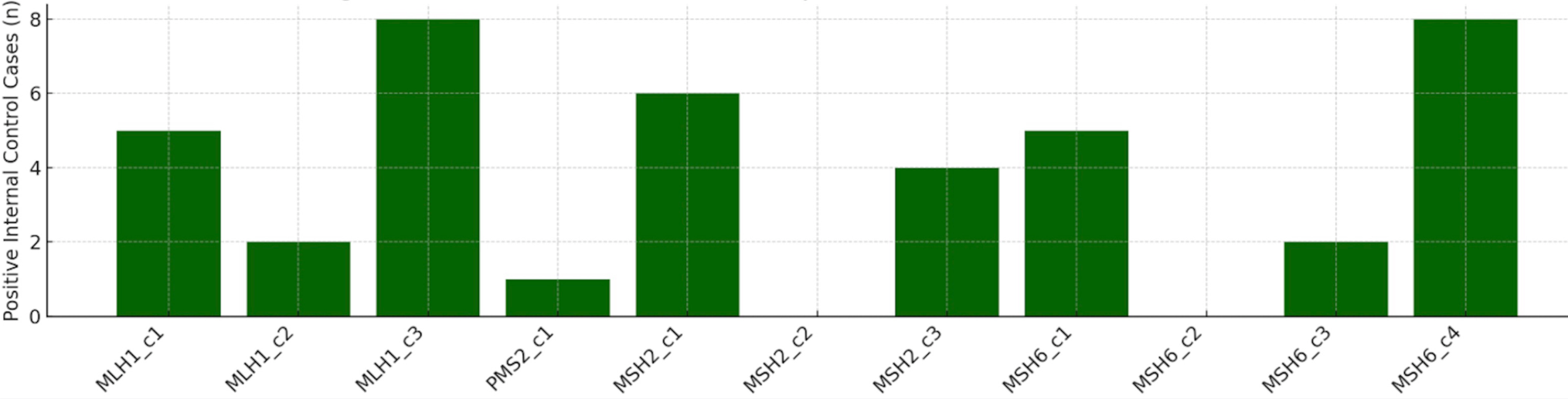
PMS2 - Only one antibody clone was available for PMS2 evaluation. This clone demonstrated weak or absent staining in normal liver tissue cores (Figure 3, Supplementary Table 3). None of the hepatocytes, cholangiocytes, fibroblasts, or inflammatory cells showed nuclear staining. In neoplastic cores, four of the six cholangiocarcinoma cores and one of the six hepatocellular carcinoma cores demonstrated preserved staining in internal control cells (Figure 4 and Figure 5). Among hepatocellular carcinomas, five tumors showed total expression loss (<25% nuclear staining) and one tumor showed intact expression (>75% nuclear staining). No cases of focal expression loss were observed. In cholangiocarcinomas, one tumor showed total expression loss (<25% nuclear staining), while five tumors demonstrated intact expression (>75% nuclear staining). No focal expression loss was detected (25%- 75%).
MSH2 - Based on control tissue staining patterns, antibody clone 3 was identified as the most effective clone (Figure 3). In normal liver tissue cores, hepatocytes showed no nuclear staining, whereas cholangiocytes, fibroblasts, and inflammatory cells demonstrated nuclear positivity for MSH2. In neoplastic tissues, both hepatocellular carcinomas and cholangiocarcinomas showed preserved staining in internal control cells (Figure 4 and Figure 5). Among hepatocellular carcinomas, two tumors demonstrated focal expression loss (25-75% nuclear staining), while two tumors showed intact expression (>75% nuclear staining). No cases of total expression loss were observed. In cholangiocarcinomas, all seven tumors demonstrated intact expression. No cases of total or focal expression loss were detected.
MSH6 - Evaluation of staining patterns in control tissues indicated that clone 4 was the most effective antibody clone (Figure 3). In normal liver tissue cores, hepatocytes, cholangiocytes, fibroblasts, and inflammatory cells all demonstrated nuclear positivity for MSH6, indicating intact expression. In neoplastic tissues, both hepatocellular carcinomas and cholangiocarcinomas showed preserved staining in internal control cells (Figure 4 and Figure 5).
Among hepatocellular carcinomas, four tumors demonstrated total expression loss (<25% nuclear staining), while four tumors showed intact expression (>75% nuclear staining). No cases of focal expression loss were observed. Among cholangiocarcinomas, all four tumors showed intact expression. No cases of total or focal expression loss were detected.
Most effective antibody clone-dependent MMR evaluation results
MMR status was further evaluated using the most effective antibody clones identified for each MMR protein. When total or focal expression loss was considered indicative of possible dMMR, the following results were observed:
For hepatocellular carcinoma, possible dMMR rates were 25% for MLH1, 50% for MSH2, and 50% for MSH6.
For cholangiocarcinoma, no cases of possible dMMR were observed for MLH1, MSH2, or MSH6.
When antibody clones other than the most effective clone were used (representing suboptimal staining conditions), the apparent frequency of possible dMMR increased due to ineffective staining (Figure 4 and Figure 5, Supplementary Table 4).
Under these conditions, possible dMMR rates ranged from 25-100% for MLH1, 100% for MSH2, and 85-100% for MSH6 in hepatocellular carcinoma.
For cholangiocarcinoma, possible dMMR rates ranged from 20-100% for MLH1, 66-75% for MSH2, and 80-100% for MSH6.
For PMS2, only one antibody clone was available, which showed suboptimal staining. Using this clone, the apparent dMMR rate was 83% for hepatocellular carcinoma and 16% for cholangiocarcinoma.
Statistical evaluation of MMR expression patterns
Based on the available Human Protein Atlas (HPA) dataset, both categorical and continuous variables were analyzed using appropriate non-parametric statistical tests.
The proportions of loss versus intact expression were compared between hepatocellular carcinoma and cholangiocarcinoma using Fisher’s exact test, whereas differences in staining intensity (0-3 scale) were assessed using the Mann-Whitney U test.
Among the MMR proteins analyzed, MSH2 demonstrated a statistically significant difference between hepatocellular carcinoma and cholangiocarcinoma (Fisher’s exact test, p = 0.015), indicating a higher frequency of expression loss in cholangiocarcinoma.
A secondary comparison of staining intensity also showed statistical significance for MSH2 (p = 0.007) and borderline significance for PMS2 (p = 0.045). No other antibodies demonstrated statistically significant differences (all p > 0.05).
Given the limited sample size and the observational nature of the HPA dataset, these findings should be interpreted as descriptive and exploratory rather than confirmatory (Figure 3, Figure 4 and Figure 5).
In supplementary analyses, comparisons of MLH1, PMS2, MSH2, and MSH6 expression levels revealed no statistically significant differences in modified H-score values among the four MMR proteins (Kruskal-Wallis test, p = 0.407), suggesting similar overall expression patterns across these markers (Supplementary Table 1).
Binary expression assessment in normal liver tissue demonstrated consistent hepatocellular and stromal staining for MLH1 and MSH6, whereas PMS2 showed minimal expression in normal liver cores (Supplementary Table 2).
Discussion
Hepatocellular carcinoma is the most common primary malignancy of the liver, followed by cholangiocarcinoma as the second most frequent type [18,19]. In this study, we focused on these two major liver tumor types and analyzed the expression status of MMR proteins using virtual slide data available in the Human Protein Atlas (HPA). This open-access platform provides a valuable resource for both pathologists and researchers by enabling systematic evaluation of immunohistochemical staining patterns across different tissues and tumor types.
Mismatch repair deficiency in hepatocellular carcinoma has been reported at varying frequencies in the literature. Bonneville et al. reported a prevalence of 0.8%, whereas Mukai et al., Kawaoka et al., and Cortes et al. reported frequencies of 2%, 2.4%, and 2.9%, respectively [4-6,20]. In the current study, higher apparent dMMR rates (25-50%) were observed compared with previously reported data. This discrepancy is most likely attributable to the limited sample size, which may not accurately represent the overall population.
For cholangiocarcinoma, MMR deficiency has been reported in 4.1% to 10% of intrahepatic cholangiocarcinomas in studies by Ju et al. and Saeed et al. [7,21]. In contrast, no dMMR cases were detected among the cholangiocarcinomas analyzed in the present study, which may similarly be explained by the limited sample size.
From an immunohistochemical perspective, MMR protein expression has been reported to be higher in normal tissues with increased proliferative activity [8]. Similarly, higher MMR expression has been observed in high-grade tumors, which typically demonstrate elevated proliferation rates [8,15]. This association between MMR expression and tumor proliferative activity may partly explain our observation of more frequent expression loss in hepatocellular carcinomas compared with cholangiocarcinomas, as the hepatocellular carcinoma cases included in the present study were predominantly low-grade tumors. This factor may also contribute to the discrepancy between the dMMR frequencies observed in this study and those reported in the literature. In addition to the limited sample size, several methodological factors may explain the higher apparent dMMR rate observed in this study. The Human Protein Atlas database does not represent a true clinical cohort and may therefore be subject to selection bias. Furthermore, MMR immunohistochemistry is highly dependent on antibody clone performance and tissue fixation quality, both of which may influence staining intensity and potentially lead to misinterpretation of reduced staining as true biological loss, particularly in well-differentiated and low-proliferation liver tumors. The use of a single tissue core per tumor may also underrepresent areas with preserved staining. Taken together, the observed dMMR frequency in this study likely reflects technical and sampling variability rather than true biological prevalence. Larger, clinicopathologically well-characterized cohorts with optimized immunohistochemical protocols and external validation are required to more accurately determine the incidence of dMMR in liver cancers.
Assessment of MMR protein expression by immunohistochemistry may present several challenges for pathologists, particularly with respect to pre-analytical variables such as tissue fixation [22]. Normal cells present within the section can serve as internal controls to help determine whether apparent loss of expression reflects fixation-related artifacts or true tumor heterogeneity [22]. However, in cases lacking an appropriate internal control, distinguishing between these possibilities becomes difficult.
Additionally, because each tumor in the HPA dataset was represented by only one or two tissue cores, the impact of intratumoral heterogeneity—particularly focal or patchy reduction of MMR protein expression—cannot be fully evaluated. In routine diagnostic practice, focal MMR alterations have been documented in several solid tumors, and sampling only one or two tumor areas may therefore underestimate or misclassify MMR status. Furthermore, in routine diagnostic pathology, the selection of tissue blocks and the choice of core sampling sites on H&E sections have a significant influence on interpretation. Preferential sampling of well-fixed, viable, and necrosis-free tumor areas improves the reliability of MMR immunohistochemistry and reduces the likelihood of artifactual staining patterns that may mimic true biological loss. Future studies incorporating multiple-core sampling and whole-slide evaluation, together with careful selection of well-preserved tumor regions on H&E sections, would better address spatial heterogeneity and help validate these observations in a more comprehensive manner.
Another important factor influencing immunohistochemical evaluation is the sensitivity of antibody clones directed against the same antigen. Differences in clone performance may account for the observed variability in staining patterns, and the use of suboptimal antibody clones can further complicate interpretation of MMR immunohistochemistry [23]. In the present study, this effect was clearly demonstrated by the significant staining differences observed among the MLH1, MSH2, and MSH6 antibody clones [23].
The number of liver cancer cases available in the Human Protein Atlas dataset was limited (n = 35), which restricts the statistical power of the analysis and precludes definitive conclusions regarding the true prevalence of MMR deficiency in hepatocellular carcinoma and cholangiocarcinoma. Therefore, the findings of this study should be interpreted as descriptive and exploratory rather than representative of the general population. In addition, each tumor was represented by a single tissue core, which may not adequately capture intratumoral heterogeneity or focal patterns of MMR protein loss. Furthermore, fixation quality and other pre-analytical conditions were not fully documented in the dataset, potentially affecting staining performance and interpretation. Future large-scale, well-annotated cohort studies incorporating comprehensive clinicopathological and molecular correlations are required to validate these observations and to better define the true frequency and clinical significance of MMR deficiency in liver malignancies.
From a clinical perspective, although the prevalence of dMMR/MSI-H in hepatocellular carcinoma and cholangiocarcinoma is low, determination of MMR status may still have important therapeutic implications. Tumors with MSI-H/dMMR across multiple organ systems are eligible for immune checkpoint inhibitor therapy, such as pembrolizumab, based on tumor-agnostic regulatory approval. Therefore, accurate interpretation of MMR immunohistochemistry is essential to avoid false-negative or false-positive assessments and to ensure appropriate patient selection for potential immunotherapy. Although routine MMR testing is not currently recommended in liver cancers because of the low frequency of dMMR, selective testing may be considered in specific clinical or pathological contexts, including young patients, unusual tumor morphology, or a strong personal or family history suggestive of MMR deficiency. Continued refinement of immunohistochemical interpretation standards and increased awareness of potential technical pitfalls will support the accurate identification of rare but clinically actionable dMMR cases.
In conclusion, this study highlights the technical complexity and interpretative challenges associated with MMR immunohistochemistry, particularly in liver pathology. Using hepatocellular carcinoma and cholangiocarcinoma as representative tumor models, we demonstrated that the interpretation of MMR staining is strongly influenced by antibody clone characteristics, tissue fixation quality, and the intrinsic proliferative and morphological features of the tumor. Well-differentiated, low-proliferative tumors such as hepatocellular carcinoma may show weak or heterogeneous staining patterns that complicate interpretation, whereas cholangiocarcinoma, with relatively higher proliferative activity, may provide clearer internal reference staining. These findings emphasize that although immunohistochemistry remains a cost-effective and widely used method for detecting dMMR, careful technical and interpretative evaluation is essential. Awareness of clone-specific staining behavior, fixation-related artifacts, and the appropriate use of internal controls is critical for accurate interpretation. Careful antibody clone selection, optimized technical protocols, and expert pathological evaluation are therefore necessary to avoid misclassification, particularly in tissues with variable fixation characteristics and low proliferative indices such as liver tumors.
Ethical approval
Ethics committee approval and informed consent were not required for this study.
Data availability statement
The data that support the findings of this study are available from the corresponding author upon reasonable request.
Conflict of interest
The author declares that this study was conducted in the absence of any commercial or financial relationships that could be construed as a potential conflict of interest.
Funding
The authors declare that this study received no funding.
Generative AI statement
The author declares that no generative AI or AI-assisted technologies were used in the writing or preparation of this study.
References
- Prolla TA, Pang Q, Alani E, Kolodner RD, Liskay RM. MLH1, PMS1, and MSH2 interactions during the initiation of DNA mismatch repair in yeast. Science 1994;265(5175):1091-3. https://doi.org/10.1126/science.8066446
- Warmerdam DO, Kanaar R. Dealing with DNA damage: relationships between checkpoint and repair pathways. Mutat Res 2010;704(1-3):2-11. https://doi.org/10.1016/j.mrrev.2009.12.001
- Vasen HF, Watson P, Mecklin JP, Lynch HT. New clinical criteria for hereditary nonpolyposis colorectal cancer (HNPCC, Lynch syndrome) proposed by the International Collaborative group on HNPCC. Gastroenterology 1999;116(6):1453-6. https://doi.org/10.1016/s0016-5085(99)70510-x
- Bonneville R, Krook MA, Kautto EA, et al. Landscape of microsatellite instability across 39 cancer types. JCO Precis Oncol 2017;2017:1-15. https://doi.org/10.1200/PO.17.00073
- Cortes-Ciriano I, Lee S, Park WY, Kim TM, Park PJ. A molecular portrait of microsatellite instability across multiple cancers. Nat Commun 2017;8:15180. https://doi.org/10.1038/ncomms15180
- Kawaoka T, Ando Y, Yamauchi M, et al. Incidence of microsatellite instability-high hepatocellular carcinoma among Japanese patients and response to pembrolizumab. Hepatol Res 2020;50(7):885-888. https://doi.org/10.1111/hepr.13496
- Ju JY, Dibbern ME, Mahadevan MS, Fan J, Kunk PR, Stelow EB. Mismatch repair protein deficiency/microsatellite instability is rare in cholangiocarcinomas and associated with distinctive morphologies. Am J Clin Pathol 2020;153(5):598-604. https://doi.org/10.1093/ajcp/aqz199
- Oshi M, Kim TH, Tokumaru Y, et al. Enhanced DNA repair pathway is associated with cell proliferation and worse survival in Hepatocellular Carcinoma (HCC). Cancers (Basel) 2021;13(2):323. https://doi.org/10.3390/cancers13020323
- Lindor NM, Burgart LJ, Leontovich O, et al. Immunohistochemistry versus microsatellite instability testing in phenotyping colorectal tumors. J Clin Oncol 2002;20(4):1043-8. https://doi.org/10.1200/JCO.2002.20.4.1043
- Zhang X, Li J. Era of universal testing of microsatellite instability in colorectal cancer. World J Gastrointest Oncol 2013;5(2):12-9. https://doi.org/10.4251/wjgo.v5.i2.12
- Batte BAL, Bruegl AS, Daniels MS, et al. Consequences of universal MSI/IHC in screening ENDOMETRIAL cancer patients for Lynch syndrome. Gynecol Oncol 2014;134(2):319-25. https://doi.org/10.1016/j.ygyno.2014.06.009
- Fadhil W, Ilyas M. Immunostaining for mismatch repair (MMR) protein expression in colorectal cancer is better and easier to interpret when performed on diagnostic biopsies. Histopathology 2012;60(4):653-5. https://doi.org/10.1111/j.1365-2559.2011.04021.x
- Shia J, Holck S, Depetris G, Greenson JK, Klimstra DS. Lynch syndrome-associated neoplasms: a discussion on histopathology and immunohistochemistry. Fam Cancer 2013;12(2):241-60. https://doi.org/10.1007/s10689-013-9612-4
- Jaffrelot M, Farés N, Brunac AC, et al. An unusual phenotype occurs in 15% of mismatch repair-deficient tumors and is associated with non-colorectal cancers and genetic syndromes. Mod Pathol 2022;35(3):427-37. https://doi.org/10.1038/s41379-021-00918-3
- The Human Protein Atlas. Available at: https://www.proteinatlas.org (Accessed on August 25, 2023).
- Uhlén M, Fagerberg L, Hallström BM, et al. Proteomics. Tissue-based map of the human proteome. Science 2015;347(6220):1260419. https://doi.org/10.1126/science.1260419
- Pontén F, Jirström K, Uhlen M. The Human Protein Atlas-a tool for pathology. J Pathol 2008;216(4):387-93. https://doi.org/10.1002/path.2440
- Pellino A, Loupakis F, Cadamuro M, et al. Precision medicine in cholangiocarcinoma. Transl Gastroenterol Hepatol 2018;3:40. https://doi.org/10.21037/tgh.2018.07.02
- Ilyas SI, Gores GJ. Pathogenesis, diagnosis, and management of cholangiocarcinoma. Gastroenterology 2013;145(6):1215-29. https://doi.org/10.1053/j.gastro.2013.10.013
- Mukai S, Kanzaki H, Ogasawara S, et al. Exploring microsatellite instability in patients with advanced hepatocellular carcinoma and its tumor microenvironment. JGH Open 2021;5(11):1266-1274. https://doi.org/10.1002/jgh3.12660
- Saeed A, Park R, Al-Jumayli M, Al-Rajabi R, Sun W. Biologics, immunotherapy, and future directions in the treatment of advanced cholangiocarcinoma. Clin Colorectal Cancer 2019;18(2):81-90. https://doi.org/10.1016/j.clcc.2019.02.005
- Torlakovic EE, Nielsen S, Francis G, et al. Standardization of positive controls in diagnostic immunohistochemistry: recommendations from the International Ad Hoc Expert Committee. Appl Immunohistochem Mol Morphol 2015;23(1):1-18. https://doi.org/10.1097/PAI.0000000000000163
- NordiQC. MMR proteins. Available at: https://www.nordiqc.org/epitope.php?id=80
Copyright and License
Copyright © 2026 The Author(s). This is an open access article distributed under the Creative Commons Attribution License (CC BY), which permits unrestricted use, distribution, and reproduction in any medium or format, provided the original work is properly cited.