


Abstract
Objectives: This study delineates the diagnostic architecture of patients referred with suspected mitochondrial disease through the integrated analysis of clinical, biochemical, instrumental, and genetic data. By comparing patients with mitochondrial involvement, alternative genetic disorders, and unresolved cases, we aim to define phenotypic and molecular patterns associated with diagnostic stratification and characterize the diverse spectrum of genetic and non-genetic conditions that converge phenotypically on mitochondrial disease.
Materials and Methods: A total of 240 patients with clinical suspicion of mitochondrial disease were consecutively enrolled and assessed using a modified Nijmegen Mitochondrial Disease Score. All participants underwent comprehensive clinical, metabolic, instrumental, and neuroimaging evaluations, complemented by systematic molecular analyses, starting with common mitochondrial variant screening and progressing to more extensive targeted investigations guided by clinical and biochemical findings. Comparative analyses across the three groups employed nonparametric and categorical statistical tests.
Results: Relevant molecular findings were identified in 81 patients (33.7%), encompassing 37 (15.4%) with mitochondrial involvement and 44 (18.3%) with alternative genetic disorders, while 159 individuals (66.3%) remained unresolved. The mitochondrial group exhibited significantly higher rates of neuromuscular, brainstem, ophthalmic, and cardiac involvement, along with developmental regression, whereas seizures were a shared hallmark of both mitochondrial and non-mitochondrial subgroups. Biochemically, elevated serum lactate and plasma alanine were the most discriminative markers for the mitochondrial group, with a significantly higher prevalence of abnormal acylcarnitine profiles and organic aciduria in both the mitochondrial and alternative genetic subgroups. The undiagnosed cohort demonstrated phenotypic convergence with confirmed cases but lacked definitive molecular correlates despite extensive evaluation.
Conclusions: These findings underscore the intrinsic complexity and phenotypic heterogeneity of suspected mitochondrial disease, affirming the critical role of integrated clinical, metabolic, instrumental, and molecular assessment while simultaneously highlighting the limitations of current diagnostic paradigms and the imperative for expanded genomic and functional strategies to enhance resolution in unresolved cases.
Keywords: mitochondrial disease, mtDNA, nijmegen mitochondrial disease score, multisystem involvement, metabolic profiling
Introduction
Mitochondrial diseases are a diverse group of inherited disorders caused by defects in mitochondrial oxidative phosphorylation, resulting in impaired cellular energy production. These conditions arise from pathogenic variants in either mitochondrial DNA (mtDNA) or nuclear genes encoding proteins essential for mitochondrial structure and function. The dual genetic origin of these disorders, together with variable heteroplasmy, the coexistence of mutant and wild-type mtDNA within the same cell, and complex genotype-phenotype relationships, contributes to marked clinical variability and a wide spectrum of disease severity, with onset ranging from childhood to adulthood [1].
Clinically, organs with high energy requirements, including the central nervous system, skeletal muscle, heart, and endocrine system, are most commonly affected. However, mitochondrial diseases lack pathognomonic clinical features and frequently share overlapping manifestations with other genetic, metabolic, and neuromuscular disorders. This substantial phenotypic overlap represents a major challenge in the diagnostic evaluation of patients with suspected mitochondrial disease and often leads to delayed, incomplete, or incorrect diagnoses [2].
The estimated prevalence of mitochondrial diseases is approximately 1 in 5,000 individuals, placing them among the most common inherited metabolic disorders. When patients with suspected but unconfirmed disease are considered, the true prevalence is likely higher, further underscoring the clinical and diagnostic burden associated with these conditions. Despite significant advances in molecular genetic techniques, a definitive diagnosis cannot be established in a considerable proportion of patients [3].
The diagnostic workup of suspected mitochondrial disease requires an integrated, multidisciplinary approach, combining clinical evaluation with biochemical, instrumental, neuroimaging, and genetic investigations. Although clinical scoring systems, such as the Nijmegen Mitochondrial Disease Score, are useful for diagnostic stratification, they lack disease specificity and may capture a broad range of non-mitochondrial conditions with overlapping phenotypes [4]. Consequently, even after extensive diagnostic assessment, many patients remain without a conclusive diagnosis.
The aim of the present study was to provide a comprehensive comparative characterization of patients with suspected mitochondrial disease, stratified by molecular outcome into: mitochondrial involvement, alternative genetic disorders, and unresolved cases despite extensive clinical and molecular evaluation. Through a multidimensional analysis of clinical, biochemical, instrumental, neuroimaging, and genetic features, we aimed to identify distinguishing profiles across these groups, to highlight the limitations of current diagnostic strategies, and to provide a framework for improving future diagnostic pathways in mitochondrial medicine.
Materials and methods
Enrollment strategy and cohort characterization
A cohort of 240 patients with clinical suspicion of mitochondrial disease was consecutively enrolled at the Institute of Mother and Child, a tertiary care hospital in the Republic of Moldova, between March 2021 and October 2024. Patients were primarily referred to our unit from various specialized departments, including neurology, cardiology, ophthalmology, and other clinical units, due to complex, multisystemic presentations that remained undiagnosed after routine investigations.
Upon referral, each patient underwent a standardized clinical assessment by our multidisciplinary team to objectively quantify the level of suspicion using a modified Nijmegen Mitochondrial Disease Score (NMDS) [5] (Table 1).
| Legend: † – indicates that this specific symptom scores 2 points; CSF – cerebrospinal fluid; MRI – magnetic resonance imaging; MRS – magnetic resonance spectroscopy. | |||
| Table 1. Modified nijmegen mitochondrial disease score | |||
| I. Clinical signs and symptoms (max. 4 points) | II. Metabolic/imaging studies (max. 4 points) |
||
| Muscular presentation (max. 2 points) |
CNS presentation (max. 2 points) |
Multisystem disease (max. 3 points) |
|
| Ophthalmoplegia † | Developmental delay | Hematology | Elevated lactate † |
| Facies myopathica | Loss of skills | Gastrointestinal tract | Elevated lactate/pyruvate ratio |
| Exercise intolerance | Stroke-like episode | Endocrine/growth Heart | Elevated alanine † |
| Muscle weakness | Migraine | Kidney | Elevated CSF lactate † |
| Rhabdomyolysis | Seizures | Vision | Elevated CSF protein |
| Abnormal electromyography | Myoclonus | Hearing | Urinary tricarbon acid excretion † |
| Cortical blindness | Neuropathy | Elevated CSF alanine † | |
| Pyramidal signs | Recurrent/familial | Ethylmalonic aciduria | |
| Extrapyramidal signs | Stroke-like picture/MRI | ||
| Brainstem involvement | Leigh syndrome/MRI † | ||
| Elevated lactate/MRS | |||
The NMDS is a validated domain-based tool that integrates clinical features, metabolic and biochemical data, and neuroimaging findings to estimate the likelihood of mitochondrial involvement. In its original form, the score also incorporates muscle biopsy and enzymatic assessments, which were not available for all patients in this cohort. To adapt to these constraints, the biopsy and enzymology domain was omitted, and the score was calculated solely based on clinical, biochemical, and neuroimaging criteria, maintaining the original scoring structure within these domains. Scores are interpreted as follows: 1 indicates mitochondrial disorder unlikely, 2-4 suggests a possible mitochondrial disorder, 5-7 corresponds to a probable mitochondrial disorder, and 8 reflects a definite mitochondrial disorder. An inclusion threshold of ≥3 points was retained to capture individuals with possible mitochondrial disease, ensuring that patients with early or partially expressed phenotypes were not excluded.
The study was approved by the Research Ethics Committee of the State University of Medicine and Pharmacy Nicolae Testemițanu and was conducted in accordance with the Declaration of Helsinki. Written informed consent was obtained from all participants or their legal guardians prior to inclusion in the study.
Patients were subsequently stratified into three categories based on diagnostic outcomes: individuals with confirmed mitochondrial involvement (n = 37), those with alternative genetic disorders (n = 44), and patients who remained without a definitive diagnosis (n = 159). Within this framework, the mitochondrial involvement group was defined by the identification of pathogenic or likely pathogenic variants, alongside specific variants of uncertain significance (VUS), including homoplasmic mtDNA mutations, that directly implicate the oxidative phosphorylation machinery. While these genetic findings provided a robust molecular explanation for most cases, this classification also encompassed patients where the detected variants offered a substantial causal contribution to the clinical presentation, serving as a foundational diagnostic anchor even in cases where they provided only a partial explanation for an exceptionally complex phenotype. Conversely, the alternative genetic disorders group was operationalized as an etiologically distinct collective originating from non-mitochondrial pathways, such as ion channelopathies or various syndromic conditions, which exhibit a marked phenotypic convergence with mitochondrial disease by replicating its multisystemic and complex clinical trajectory. Finally, the unresolved group comprised individuals who met the inclusion criteria but in whom no causative genetic variants were identified, representing the inherent diagnostic limitations in current genomic screening. In the following sections, we detail our institutional experience across these three cohorts, providing a comprehensive appraisal of the clinical, biochemical, and molecular patterns observed to improve the differential diagnosis between confirmed or suspected mitochondrial involvement and its genetic mimics.
Clinical, familial, and paraclinical evaluation
All participants underwent a thorough clinical assessment, including detailed medical history, family history, prenatal and perinatal data, and age at symptom onset. Systematic physical and neurological examinations were conducted to evaluate multisystem involvement. Routine laboratory testing encompassed hematological indices, renal and hepatic function, serum electrolytes, lactate, creatine kinase, and lactate dehydrogenase.
Extended metabolic investigations, conducted for a subset of the cohort, encompassed plasma amino acid profiling via high-performance liquid chromatography, acylcarnitine quantification by liquid chromatography-tandem mass spectrometry, and urinary organic acid analysis using nuclear magnetic resonance spectroscopy. Pathological status was defined by persistent deviations from laboratory-established reference intervals, encompassing disproportionate accumulations of short-, medium-, and long-chain acylcarnitine species, aberrant diagnostic ratios indicative of impaired intermediary metabolism, or the abnormal excretion of organic acid biomarkers, including dicarboxylic, methylmalonic, and glutaric acids, alongside elevated Krebs cycle intermediates.
Instrumental and imaging studies were performed based on clinical indications, comprising electroencephalography, electromyography, electrocardiography, audiometry, and cerebral imaging (MRI or CT) to detect structural or functional abnormalities.
Genetic analysis
All 240 patients underwent initial molecular prescreening for seven recurrent pathogenic mtDNA point mutations, specifically m.3243A>G, m.8344A>G, m.8993T>G/C, m.13513G>A, m.3460G>A, m.11778G>A, and m.14484T>C, using quantitative polymerase chain reaction with high-resolution melting analysis (qPCR-HRM). To ensure diagnostic precision and calibrate melting profiles, synthetic oligonucleotide controls representing both wild-type and mutant genotypes were systematically employed. Heteroplasmy levels for variants identified via qPCR-HRM were further evaluated using PCR-restriction fragment length polymorphism, with semi-quantitative assessment performed through densitometric analysis of electrophoresis gels using ImageJ software.
Patients with a modified NMDS ≥6, as well as those exhibiting inconclusive results in the prescreening, were advanced to targeted Sanger sequencing of the mitochondrial genome. This threshold of ≥6 was strategically established to prioritize molecular resources for a subgroup with the highest pre-test probability of mitochondrial disease. Within the NMDS framework, a score exceeding 6 ensures that clinical signs (which contribute a maximum of 4 points in Category I) are supported by objective findings from Category II, such as hyperlactatemia, hyperalaninemia, or specific mitochondrial signatures on brain MRI. The analysis covered all 13 protein-coding genes and included flanking tRNA and rRNA genes adjacent to the coding regions, ensuring comprehensive assessment of loci relevant to mitochondrial disease. Detected variants were interpreted according to the American College of Medical Genetics and Genomics (ACMG) guidelines [6], ensuring a standardized and clinically meaningful classification framework.
A subset of patients underwent additional molecular analyses of the nuclear genome using targeted Sanger sequencing and next-generation sequencing (NGS) technologies, including whole-exome sequencing (WES), whole-genome sequencing (WGS) and targeted multigene panels. These genomic investigations were complemented in some instances by Multiplex Ligation-dependent Probe Amplification (MLPA) or array comparative genomic hybridization (aCGH). All procedures were conducted in ISO 15189-accredited laboratories, both domestically and internationally, ensuring methodological rigor, reproducibility, and adherence to recognized quality standards. These comprehensive analyses facilitated the identification of nuclear gene variants implicated in mitochondrial dysfunction, as well as the detection of variants unrelated to mitochondrial disease. Detected variants were systematically interpreted and classified according to the ACMG guidelines, considering pathogenicity, allele frequency, predicted functional impact, and available clinical evidence, thereby providing a standardized and clinically meaningful framework for molecular diagnosis.
DNA extraction for all molecular investigations, including qPCR-HRM screening, Sanger sequencing, and NGS-based analyses, was performed exclusively from peripheral blood samples.
Statistical analysis
Statistical analyses were performed using SPSS version 28.0 (IBM Corp., USA). Continuous variables were expressed as mean ± standard deviation or median with interquartile range, whereas categorical variables were summarized as counts and percentages. Comparisons across the three investigation groups were conducted using the Kruskal-Wallis test for continuous variables and the Chi-square test for categorical variables. A significance threshold of p < 0.05 was applied.
Results
The study population comprised 240 patients with suspected mitochondrial disease, stratified into three groups based on molecular outcomes: 37 (15.4%) with mitochondrial involvement, 44 (18.3%) with alternative genetic disorders, and 159 patients (66.3%) remained without a confirmed diagnosis.
The median age at study inclusion was 24 months (IQR: 11–68) for the mitochondrial involvement group, 24 months (IQR: 12–72) for alternative genetic disorders, and 30 months (IQR: 11–66) in the undiagnosed group, indicating substantial overlap in age distribution between the cohorts. Symptom onset occurred predominantly in early infancy, with a median age of 3 months (IQR: 1–15) in the mitochondrial group, 4 months (IQR: 1–10) in the alternative disorders group, and 2 months (IQR: 0–12) in undiagnosed patients.
No statistically significant differences were observed across the three investigation groups with respect to the presence of a positive family history of similar clinical manifestations. Additionally, the frequency of pregnancy and birth-related complications, including preterm delivery, intrauterine growth restriction, and other perinatal adversities, was comparable across groups.
Clinical manifestations
The clinical profile of the study cohort was highly heterogeneous, reflecting the multisystemic nature of suspected mitochondrial dysfunction. This diversity provided a structured framework for a comparative analysis of clinical prevalence and severity across the three investigation groups, with the distribution of these features among patients with mitochondrial involvement, alternative genetic disorders, and unresolved cases illustrated in Figure 1.
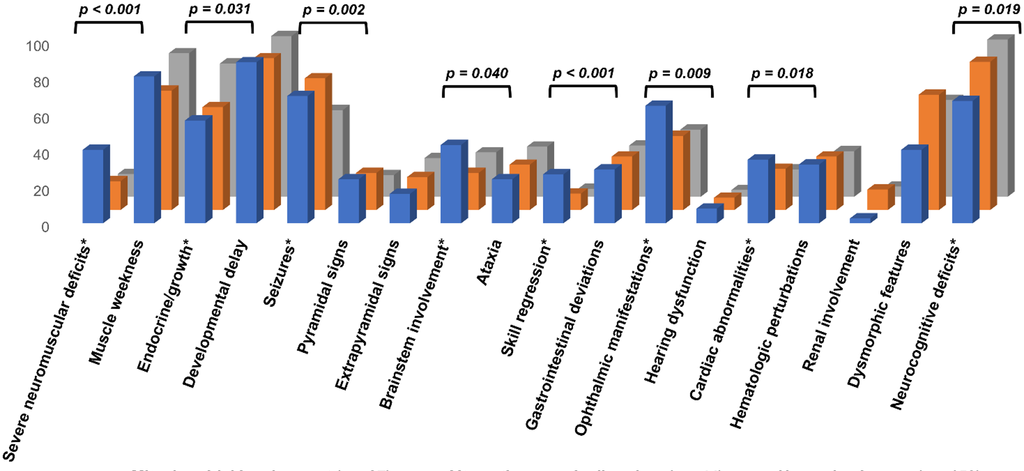
The mitochondrial involvement group exhibited significantly higher rates of severe neuromuscular deficits (40.5% vs. 15.9% and 12.6%; p < 0.001), brainstem involvement (43.2% vs. 20.5% and 24.5%; p = 0.040), ophthalmic manifestations (64.9% vs. 40.9% and 37.1%; p = 0.009), cardiac abnormalities (35.1% vs. 22.7% and 15.1%; p = 0.018), and skill regression (27.0% vs. 9.1% and 4.4%; p < 0.001) compared to the alternative genetic disorders and unresolved groups, respectively. Seizures were a shared predominant feature in both the mitochondrial involvement (70.3%) and alternative genetic disorders (72.7%) groups, occurring significantly more frequently than in the unresolved cohort (47.8%; p = 0.002).
Conversely, undiagnosed patients exhibited a higher prevalence of neurodevelopmental and behavioral impairments (86.8% vs. 67.6% and 81.8%; p = 0.021) endocrine or growth-related abnormalities (73.6% vs. 56.8% and 56.8%; p = 0.011), relative to the mitochondrial involvement and alternative genetic disorders groups, respectively.
Biochemical and instrumental investigations
Biochemical testing revealed that elevated serum lactate emerged as a discriminative marker between groups, being both significantly more prevalent as an abnormal finding in patients with mitochondrial involvement (34/37, 91.9%) compared to both the alternative genetic disorders (16/38, 42.1%) and unresolved (57/138, 41.3%) cohorts. Furthermore, a comparison of absolute concentrations using the Kruskal-Wallis test demonstrated that lactate levels were significantly higher in the mitochondrial group (Mean Rank: 159.6) than in the alternative (Mean Rank: 95.0) and unresolved (Mean Rank: 96.2) groups (H = 32.66, p < 0.001). Plasma alanine demonstrated a similar discriminative pattern, with significant differences observed in both prevalence (p < 0.001) and absolute concentrations across the three investigation groups (H = 22.92, p < 0.001). This significance was primarily driven by the mitochondrial involvement group, which presented elevated alanine levels in 60.0% (18/30) of patients, substantially exceeding the frequencies observed in the alternative genetic disorders (23.5%, 8/34) and unresolved (11.2%, 13/116) cohorts (Mean Ranks: 87.1, 59.0, and 46.5, respectively).
Additional metabolic screening identified significant disparities in secondary markers of metabolic dysfunction, revealing that pathological acylcarnitine profiles (p = 0.027) were notably more frequent in the alternative genetic disorders group (32.3%, 10/31) compared to the mitochondrial involvement (25.0%, 5/20) and unresolved (12.7%, 15/118) cohorts. Furthermore, while infrequent across all groups, organic aciduria (p = 0.008) was more common in the mitochondrial involvement (16.7%, 5/30) and alternative genetic disorders (13.5%, 5/37) groups than in the unresolved cohort (3.1%, 4/130).
Other routine laboratory parameters, including serum transaminases, creatine kinase, lactate dehydrogenase, and serum electrolytes, did not differ significantly between groups. The distribution of biochemical, instrumental, and neuroimaging abnormalities across the mitochondrial involvement, alternative genetic disorders, and unresolved groups is summarized in Figure 2.
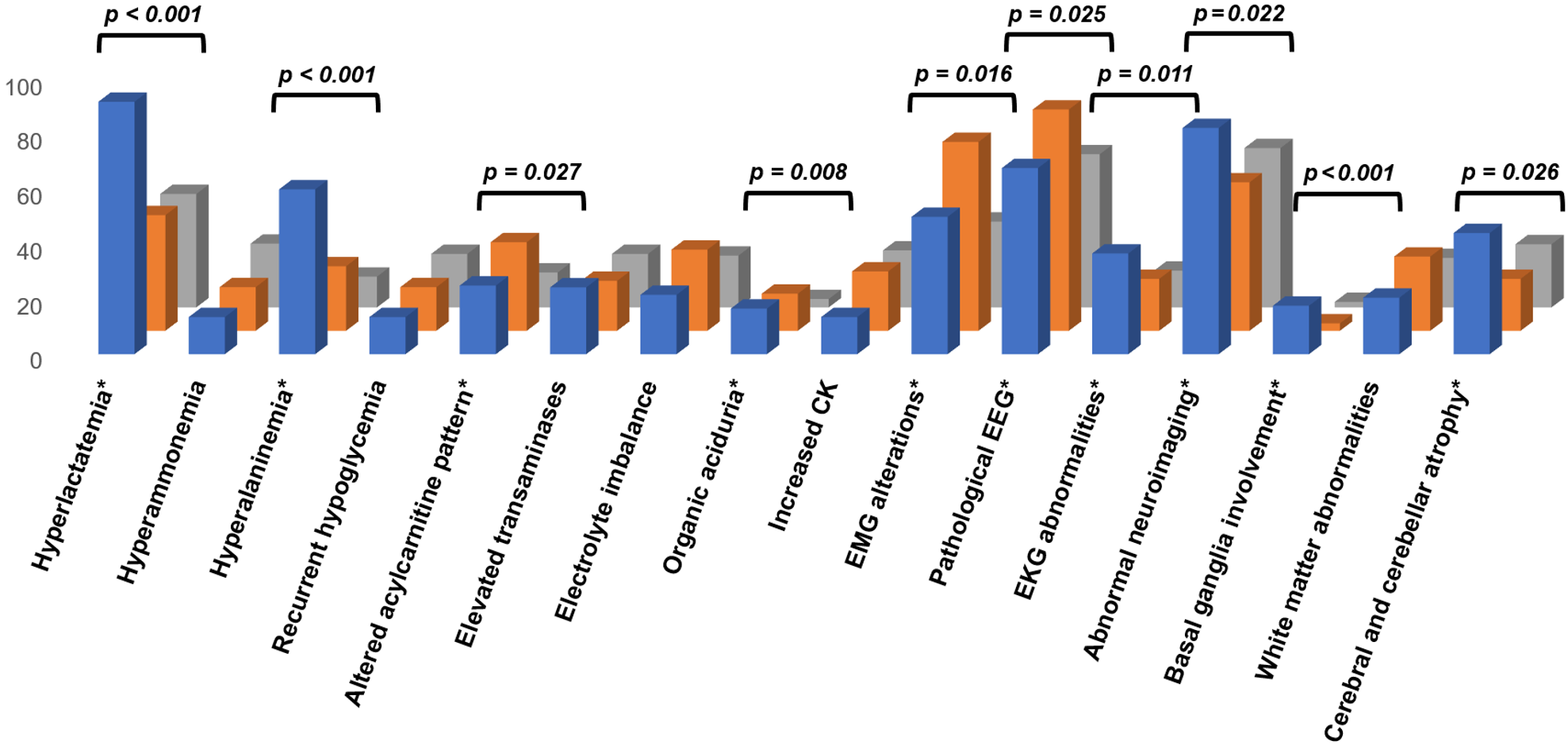
Instrumental assessments revealed marked distinctions between cohorts. Electromyographic abnormalities reached their highest frequency in the alternative genetic disorders group (68.8%, 11/16), followed by the mitochondrial involvement group (50.0%, 9/18), with both cohorts showing significantly higher rates than the 31.3% (20/64) identified in the unresolved cohort (p = 0.016). This was paralleled by a higher frequency of electroencephalographic alterations in the alternative genetic (80.6%, 29/36) and mitochondrial (67.7%, 21/31) groups compared to unresolved cases (55.8%, 58/104; p = 0.025), alongside cardiographic deviations which were notably more prevalent in the mitochondrial involvement group (36.7%, 11/30) relative to the alternative genetic (18.9%, 7/37) and unresolved (13.4%, 17/127) cohorts (p = 0.011).
Neuroimaging abnormalities were significantly more prevalent in the mitochondrial involvement group (82.4%, 28/34) compared to the alternative genetic disorders (54.1%, 20/37) and unresolved (58.0%, 58/100) cohorts (p = 0.022). Specifically, the mitochondrial involvement group exhibited higher rates of cerebral or cerebellar atrophy (44.1% vs. 18.9% and 23.0%; p = 0.026) and basal ganglia involvement (17.6% vs. 2.7% and 2.0%; p < 0.001), underscoring a distinct neuroanatomical pattern of mitochondrial pathology.
The cumulative burden of these multi-systemic findings was further reflected in the clinical stratification provided by the NMDS. Within the mitochondrial involvement group, clinical suspicion was most intense, with 10 patients reaching a definite mitochondrial disease classification and 26 a probable one, compared to only 1 possible case. In the alternative genetic disorders group, the distribution shifted toward milder clinical features, involving 3 definite, 17 probable, and 24 possible cases. In contrast, the unresolved cohort was heavily skewed toward lower clinical suspicion, being dominated by 98 possible and 59 probable qualifiers, with only 2 patients meeting the definite mitochondrial disease criteria. Ultimately, significantly higher NMDS scores were concentrated in the mitochondrial group (p < 0.001), confirming the scale’s effectiveness in identifying individuals whose multi-organ clinical burden strongly aligns with a mitochondrial-related phenotype.
Genetic investigations
Genetic evaluation contributed decisively to case stratification within the study population. All patients underwent initial molecular screening using a qPCR-HRM-based approach targeting common pathogenic mtDNA variants. Through this first-line screening, eight patients were identified as carriers of established pathogenic mtDNA point mutations. Specifically, the m.3243A>G variant was detected in three patients, m.8993T>G in two patients, m.3460G>A in two patients, and m.11778G>A in one patient, all of whom were classified as molecularly confirmed mitochondrial cases.
Subsequently, a total of 82 patients with increased clinical suspicion, defined by an NMDS ≥6, underwent extended molecular analysis of the mitochondrial genome by targeted Sanger sequencing with capillary electrophoresis. This approach led to the identification of 24 additional patients harboring mtDNA variants: 11 individuals (45.8%) with variants classified as pathogenic or likely pathogenic and 13 patients (54.2%) with VUS. Although these VUS cases do not fulfill the criteria for formal molecular confirmation, they were included in the mitochondrial involvement group based on the presence of homoplasmic variants that closely correlated with the specific clinical phenotypes of the patients. Analysis of the functional distribution of these variants revealed a predominant impact on respiratory chain architecture, with mutations affecting Complex I in 25.0% of cases, followed by alterations involving Complex V in 12.5% and mitochondrial RNA genes in 16.7%. In contrast, variants affecting Complexes III and IV were comparatively infrequent, each accounting for 4.1% of cases, while a substantial proportion of patients (37.6%) harbored variants predicted to compromise multiple respiratory chain complexes, underscoring the extensive and functionally heterogeneous nature of mitochondrial genomic involvement within this clinically selected subgroup.
In selected patients presenting with sustained clinical features suggestive of mitochondrial involvement, extended nuclear genetic testing was undertaken to further elucidate the underlying molecular etiology. This diagnostic effort, utilizing targeted Sanger sequencing (n = 2) and WES (n = 3), yielded significant findings in five individuals. These investigations identified pathogenic or likely pathogenic variants in POLG (n = 2), TWNK, and ETHE1, alongside a VUS in the DGUOK gene. Notably, one patient evaluated via WES demonstrated dual molecular involvement, harboring a pathogenic mtDNA alteration, previously detected by qPCR-HRM, concomitant with a VUS in the OPA1 gene.
Altogether, the integration of these findings resulted in a total of 37 patients with molecular evidence of mitochondrial involvement, encompassing both mitochondrial and nuclear genetic determinants. Detailed profiles of the identified genetic variants and corresponding patient phenotypes are summarized in Supplementary Table 1.
Among patients with non-mitochondrial conditions, genetic testing identified alternative molecular diagnoses in 44 cases, comprising 32 individuals with pathogenic or likely pathogenic variants and 12 with VUS. The diagnostic workflow utilized a multimodal approach primarily leveraging high-throughput sequencing (WGS, n = 15; WES, n = 14; targeted multigene panels, n = 6), supplemented by structural variant analysis (MLPA, n = 4; aCGH, n = 1), and targeted Sanger sequencing (n = 4). These findings were distributed across five principal diagnostic categories. Syndromic and sensory disorders represented the largest group (n = 15, 34.1%), with variants in genes associated with neurodevelopmental and multisystem syndromes, including FOXG1, NSD1, PPP2R5D and others. The inborn errors of metabolism group (n = 12, 27.3%) involved defects in amino acid, carbohydrate, lipid, and peroxisomal metabolism, exemplified by variants in GLB1, PCCA, ALDOB, and additional genes. Channelopathies (n = 8, 18.1%) were associated with variants in voltage-gated ion channel genes such as SCN1A, SCN2A, and SCN10A. Neuromuscular disorders (n = 4, 9.1%) included variants in ANO5, DARS2, and TCAP, affecting muscle integrity and protein translation. Finally, chromosomal microdeletion syndromes (n = 5, 11.4%), encompassed recurrent pathogenic regions including 4p16.3, 7q11.23, and 15q11–q13, among others, further illustrating the broad spectrum of genetic conditions that can phenotypically mimic suspected mitochondrial disease. The molecular characteristics of the identified variants and their associated clinical phenotypes are systematically presented in Supplementary Table 2.
Patients who remained without a definitive molecular diagnosis were subjected to the same initial qPCR-HRM screening; however, no pathogenic mtDNA variants fulfilling diagnostic criteria were detected, and further genetic evaluations, encompassing the targeted analyses employed in this study, did not yield conclusive molecular evidence, underscoring the limitations of current methodologies in resolving these complex cases.
Discussion
We evaluated a pediatric cohort with suspected mitochondrial disease by integrating clinical, biochemical, instrumental, and genetic data. By comparatively analyzing patients across the categories of confirmed mitochondrial involvement, alternative genetic disorders, and unresolved cases, our findings pinpoint specific phenotypic patterns that improve patient classification and reflect the diverse nature of mitochondrial pathology.
Despite broadly comparable demographic and perinatal characteristics, patients with mitochondrial involvement exhibited a markedly more severe and multisystemic phenotype. This subgroup demonstrated higher rates of severe neuromuscular dysfunction, brainstem involvement, ophthalmic manifestations, cardiovascular abnormalities, and developmental regression. These clinical features reflect the heightened susceptibility of high-energy-dependent tissues to mitochondrial impairment and confirm that extensive multisystemic involvement remains a key indicator of mitochondrial disease [7-8]. Seizures were a predominant feature in both the mitochondrial involvement and alternative genetic disorders groups. Conversely, patients lacking molecular confirmation were predominantly distinguished by neurodevelopmental, behavioral, and growth-related abnormalities, which, while often prompting suspicion of mitochondrial dysfunction, are more frequently associated with alternative genetic etiologies exhibiting overlapping phenotypic features.
Biochemical profiling further distinguished these groups, with elevated serum lactate and plasma alanine emerging as the most discriminative metabolic markers for the mitochondrial involvement group, indicative of impaired oxidative phosphorylation and secondary perturbations in intermediary metabolism. Pathological acylcarnitine profiles and organic aciduria were more frequently observed in both the mitochondrial involvement and alternative genetic disorders subgroups. These findings highlight the importance of expanded biochemical screening for identifying both the metabolic effects of mitochondrial failure and other underlying systemic metabolic abnormalities.
The Nijmegen Mitochondrial Disease Score demonstrated strong utility in capturing multisystem involvement, with significantly higher values in the mitochondrial involvement group (p < 0.001). Given that relatively high scores were also noted in the alternative genetic group, in our setting, the NMDS effectively identified cases more likely to yield relevant molecular findings across the entire cohort.
Comprehensive genetic analyses, encompassing both targeted mitochondrial and selected nuclear testing, identified relevant molecular findings in 33.7% of patients. This approach effectively stratified patients according to molecular etiology and highlighted the critical role of integrated genomic evaluation in the diagnostic workflow of suspected mitochondrial disease. Within this context, the diagnostic distribution observed in the present study is concordant with that reported in previous investigations of large and medium-sized cohorts of patients referred with suspected mitochondrial disease, which consistently demonstrate a heterogeneous composition including confirmed mitochondrial disorders, alternative non-mitochondrial genetic conditions, and a substantial proportion of unresolved cases. In the largest series reported to date, Rouzier et al. [9] analyzed a cohort exceeding 2,000 patients and established a molecular diagnosis in approximately 20% of cases, identifying 322 patients with mitochondrial disease and 75 with non-mitochondrial genetic etiologies, while the majority of individuals remained without a definitive diagnosis. Similarly, van der Ven et al. [10] investigated 491 patients and achieved a diagnostic yield of 51% (n = 250), of whom 221 patients (45%) were diagnosed with non-mitochondrial disorders and 29 patients (6%) with mitochondrial disease, whereas 241 patients (49%) remained undiagnosed. Comparable results were reported by Kerr et al. [11], who evaluated 390 patients and established a molecular diagnosis in 184 cases (47.2%), including 115 patients (29.5%) with non-mitochondrial genetic disorders and 69 patients (17.7%) with mitochondrial disease, with 206 patients (52.8%) remaining without molecular resolution. At the family-based level, Schon et al. [12] analyzed 319 families and identified a genetic diagnosis in 104 families (32.6%), comprising 65 families (20.4%) with non-mitochondrial causes and 39 families (12.2%) with mitochondrial disease, while 215 families (67.4%) remained without a confirmed molecular diagnosis. In smaller cohorts, such as that reported by Grigalionienė et al. [13], diagnostic yields were similarly limited; among 83 patients, only 18 (21.7%) received a molecular diagnosis, including 11 patients (13.3%) with mitochondrial disease, six patients (7.2%) with non-mitochondrial disorders, and one patient (1.2%) with combined mitochondrial and other genetic pathology, leaving 65 patients (78.3%) undiagnosed. Collectively, these data place our findings within a robust and reproducible diagnostic paradigm characteristic of suspected mitochondrial disease.
While this study provides a comprehensive evaluation, several methodological limitations merit consideration when interpreting the findings. Although the cohort benefited from extensive clinical, biochemical, and instrumental characterization, invasive functional investigations, most notably muscle biopsy with histopathological, histochemical, and respiratory chain enzymatic analyses, could not be systematically performed, precluding direct tissue-level confirmation of mitochondrial dysfunction and potentially limiting diagnostic resolution in selected cases. Furthermore, genetic investigations were conducted exclusively using peripheral blood samples. While this non-invasive approach is standard for primary screening and provides adequate DNA yield, it may underrepresent tissue-specific heteroplasmy or fail to detect certain mitochondrial DNA alterations that are more reliably identified in post-mitotic tissues, such as muscle. Additionally, the use of Sanger sequencing for mtDNA analysis precluded the precise quantification of heteroplasmy levels; however, the majority of variants we identified appeared homoplasmic, aligning with the expected molecular profile for these specific alterations.
Moreover, although the genetic diagnostic strategy was structured and clinically guided, it relied primarily on targeted assays, and the limited use of NGS constrained comprehensive interrogation of both nuclear and mitochondrial genomes. This methodological constraint is especially relevant considering that expanded NGS approaches, including whole exome and dual genome analyses, have been shown to substantially increase the diagnostic yield in suspected mitochondrial disorders [14-16]. In our setting, the restricted application of these advanced technologies reflects persistent regional challenges; as high-throughput genomic testing is not yet integrated into the national healthcare reimbursement system, its implementation remains largely dependent on the financial resources of the patients’ families. Consequently, pathogenic variants beyond the scope of the applied methods may have remained undetected, contributing to the persistence of unresolved cases. Finally, the pediatric, referral-based nature of the cohort may constrain the generalizability of these findings to adult populations or unselected clinical settings. Nonetheless, these constraints mirror real-world diagnostic practice and highlight the continued need for integrated functional approaches and broader genomic strategies to improve diagnostic resolution in patients with suspected mitochondrial disease.
Conclusion
This study delivers a rigorous, multidimensional characterization of a pediatric cohort with suspected mitochondrial disease, integrating clinical, biochemical, instrumental, and genetic data to refine diagnostic stratification. Relevant molecular findings were identified in 33.7% of patients (n = 81), distributed between patients with mitochondrial involvement (n = 37) and alternative genetic disorders (n = 44), while 66.3% (n = 159) remained unresolved. Specifically, patients with mitochondrial involvement demonstrated pronounced multisystemic involvement, distinctive metabolic perturbations, and convergent neuroanatomical alterations, reflecting the complex pathophysiology and systemic impact of mitochondrial dysfunction. The results underscore the critical importance of integrated diagnostic frameworks that combine detailed clinical assessment with targeted and expanded genomic analyses. Beyond current strategies, there remains a clear necessity for advanced sequencing technologies and functional investigations to enhance diagnostic resolution and support precision management in complex clinical settings.
Ethical approval
This study was approved by the Research Ethics Committee of the State University of Medicine and Pharmacy “Nicolae Testemițanu” (Date: September 9, 2020, Decision/Protocol No: 03). Informed consent was obtained from all participants involved in this study.
Data availability statement
The data that support the findings of this study are available from the corresponding author upon reasonable request.
Conflict of interest
The authors declare that this study was conducted in the absence of any commercial or financial relationships that could be construed as a potential conflict of interest.
Funding
This study was supported by the institutional research project “Diagnosis and Monitoring of Genetic Diseases in the Prevention of Maternal and Child Health Disorders” (DiMoGEN, 140102) and by the project “Genomic Medicine and Metabolomics Research in the Service of Prophylaxis of Genetic Diseases for Healthy Generations in the Republic of Moldova” (SCREENGEN, 20.80009.8007.22).
Generative AI statement
The authors declare that during the preparation of this study, the following AI-assisted technology was used: ChatGPT-5.5 on December 2025 - January 2026. Extent of Use: The tool was used exclusively for English language editing, proofreading, and improving the phrasing and grammatical correctness of the manuscript. It was not used to generate any new scientific content, ideas, or data analysis. The authors confirm that they have critically reviewed and edited any AI-generated content and take full responsibility for the integrity, accuracy, and originality of the publication. The authors certify that the original human contribution is maintained and that AI-assisted tools are not listed or cited as authors.
References
- Hong S, Kim S, Kim K, Lee H. Clinical approaches for mitochondrial diseases. Cells 2023;12(20):2494. https://doi.org/10.3390/cells12202494
- Grier J, Hirano M, Karaa A, Shepard E, Thompson JLP. Diagnostic odyssey of patients with mitochondrial disease: results of a survey. Neurol Genet 2018;4(2):e230. https://doi.org/10.1212/NXG.0000000000000230
- Wen H, Deng H, Li B, et al. Mitochondrial diseases: from molecular mechanisms to therapeutic advances. Signal Transduct Target Ther 2025;10(1):9. https://doi.org/10.1038/s41392-024-02044-3
- Forny P, Footitt E, Davison JE, et al. Diagnosing mitochondrial disorders remains challenging in the omics era. Neurol Genet 2021;7(3):e597. https://doi.org/10.1212/NXG.0000000000000597
- Morava E, van den Heuvel L, Hol F, et al. Mitochondrial disease criteria: diagnostic applications in children. Neurology 2006;67(10):1823-6. https://doi.org/10.1212/01.wnl.0000244435.27645.54
- Richards S, Aziz N, Bale S, et al. Standards and guidelines for the interpretation of sequence variants: a joint consensus recommendation of the American College of Medical Genetics and Genomics and the Association for Molecular Pathology. Genet Med 2015;17(5):405-24. https://doi.org/10.1038/gim.2015.30
- Di Donato S. Multisystem manifestations of mitochondrial disorders. J Neurol 2009;256(5):693-710. https://doi.org/10.1007/s00415-009-5028-3
- Klopstock T, Priglinger C, Yilmaz A, Kornblum C, Distelmaier F, Prokisch H. Mitochondrial disorders. Dtsch Arztebl Int 2021;118(44):741-8. https://doi.org/10.3238/arztebl.m2021.0251
- Rouzier C, Pion E, Chaussenot A, et al. Primary mitochondrial disorders and mimics: Insights from a large French cohort. Ann Clin Transl Neurol 2024;11(6):1478-91. https://doi.org/10.1002/acn3.52062
- van der Ven AT, Johannsen J, Kortüm F, et al. Prevalence and clinical prediction of mitochondrial disorders in a large neuropediatric cohort. Clin Genet 2021;100(6):766-70. https://doi.org/10.1111/cge.14061
- Kerr M, Hume S, Omar F, et al. MITO-FIND: a study in 390 patients to determine a diagnostic strategy for mitochondrial disease. Mol Genet Metab 2020;131(1-2):66-82. https://doi.org/10.1016/j.ymgme.2020.08.009
- Schon KR, Horvath R, Wei W, et al. Use of whole genome sequencing to determine genetic basis of suspected mitochondrial disorders: cohort study. BMJ 2021;375:e066288. https://doi.org/10.1136/bmj-2021-066288
- Grigalionienė K, Burnytė B, Ambrozaitytė L, Utkus A. Wide diagnostic and genotypic spectrum in patients with suspected mitochondrial disease. Orphanet J Rare Dis 2023;18(1):307. https://doi.org/10.1186/s13023-023-02921-0
- Gorman E, Dai H, Feng Y, et al. Experiences from dual genome next-generation sequencing panel testing for mitochondrial disorders: a comprehensive molecular diagnosis. Front Genet 2025;16:1488956. https://doi.org/10.3389/fgene.2025.1488956
- Mahmud S, Biswas S, Afrose S, et al. Use of next-generation sequencing for identifying mitochondrial disorders. Curr Issues Mol Biol 2022;44(3):1127-48. https://doi.org/10.3390/cimb44030074
- Olimpio C, Paramonov I, Matalonga L, et al. Increased diagnostic yield by reanalysis of whole exome sequencing data in mitochondrial disease. J Neuromuscul Dis 2024;11(4):767-75. https://doi.org/10.3233/JND-240020
Copyright and License
Copyright © 2026 The Author(s). This is an open access article distributed under the Creative Commons Attribution License (CC BY), which permits unrestricted use, distribution, and reproduction in any medium or format, provided the original work is properly cited.